


Lamellar procedures have their origins in the 1960s, when they were introduced by Barraquer in Columbia.1 In the 1990s, Pallikaris and Buratto introduced the concept of combining a lamellar procedure with surface ablation excimer lasers,2,3 giving rise to laser-assisted in situkeratomileusis (LASIK) surgery. Several iterations and techniques have been developed since then, and over the last three decades LASIK has become one of the most common and successful elective refractive procedures performed.4,5 Since its inception, more than 15 million patients have undergone the surgery.6 Overall, the complication rate from LASIK is low, with reports ranging from <1–1.8%.7–9
层状手术起源于 1960 年代,当时它们由哥伦比亚的 Barraquer 引入。在 1990 年代,Pallikaris 和 Buratto 引入了将层状手术与表面消融准分子激光相结合的概念, 2,3 从而产生了激光辅助原位肌位 (LASIK) 手术。从那时起,已经开发了几种迭代和技术,在过去的三十年中,LASIK已成为最常见和最成功的选择性屈光手术之一。 4,5 自成立以来,已有超过 1500 万患者接受了手术。 6 总体而言,LASIK的并发症发生率较低,报告范围为<1-1.8%。 7–9
The first critical step in LASIK is creation of the corneal flap. The LASIK flap is paramount to the procedure and allows for quick visual recovery with little pain,10 but is also a major source of complications. There are two main methods for creating corneal flaps. The first involves using a mechanical microkeratome with an oscillating blade.11 The second utilizes a femtosecond laser with a focusable, infrared-spectrum photodisruptive laser, which forms cavitation bubbles that spread to produce a dissected corneal flap.12
LASIK的第一个关键步骤是角膜瓣的创建。LASIK皮瓣对手术至关重要,可以快速恢复视力,几乎没有疼痛, 10 但也是并发症的主要来源。创建角膜瓣有两种主要方法。第一种涉及使用带有摆动刀片的机械微型角膜刀。 11 第二种利用飞秒激光和可聚焦的红外光谱光干扰激光,形成空化气泡,扩散产生解剖的角膜瓣。 12
Advantages of femtosecond laser flap creation include reduced variation in flap thickness and increased repeatability.13 Some studies have suggested better final visual acuity,14 lower intraocular pressure (IOP) during the flap creation,15 and lower incidence of dry eye.16 Additionally, some prominent microkeratome complications including free/incomplete flaps, buttonholes, and epithelial erosions are less common due to the femtosecond’s precision.14 However, disadvantages of the femtosecond include transient light sensitivity,17 diffuse lamellar keratitis,18 rainbow glare,19 opaque bubble layer,20 and cost.21 Both device classes are still used, but femtosecond has grown in popularity over time. Several femtosecond laser systems are approved by The Food and Drug Administration (FDA) for LASIK use. However, discussion on each individual system is beyond the scope of this review.
创建飞秒激光襟翼的优点包括减少襟翼厚度的变化和提高可重复性。 13 一些研究表明,最终视力更好, 14 皮瓣制作过程中的眼压 (IOP) 更低, 15 干眼症的发生率更低。 16 此外,由于飞秒的精度,一些突出的显微角膜刀并发症,包括游离/不完全皮瓣、扣眼和上皮糜烂,不太常见。 14 然而,飞秒的缺点包括瞬态光敏感、 17 弥漫性层状角膜炎、 18 彩虹眩光、 19 不透明气泡层 20 和成本。 21 这两种设备类别仍在使用,但飞秒随着时间的推移越来越受欢迎。美国食品和药物管理局 (FDA) 批准了几种飞秒激光系统用于 LASIK。但是,对每个系统的讨论超出了本综述的范围。
Understanding, avoiding and managing potential femtosecond flap complications is essential for anyone performing femtosecond-assisted LASIK. This review will discuss intraoperative and postoperative flap complications associated with femtosecond-assisted LASIK using PubMed-identified relevant published literature. The PubMed search was conducted using “LASIK flap complications” as a primary search term, as well as “LASIK” alongside the various associated complications that will be discussed later.
了解、避免和管理潜在的飞秒皮瓣并发症对于任何进行飞秒辅助LASIK手术的人来说都是必不可少的。本综述将使用PubMed确定的相关已发表文献讨论与飞秒辅助LASIK相关的术中和术后皮瓣并发症。PubMed检索使用“LASIK皮瓣并发症”作为主要检索词,以及“LASIK”以及稍后将讨论的各种相关并发症。
Intraoperative complications
术中并发症
Suction loss 吸力损失
While not unique to femtosecond flap creation, issues with suction are one of the most commonly encountered problems. Reported rates of suction loss range from 0.06–0.8%.22 Given the unique lamellar cutting ability of femtosecond lasers, suction issues can often be overcome without compromising patient outcomes.23 Unlike keratome flaps, procedures are not automatically aborted when suction loss occurs.4
虽然飞秒皮瓣的产生并非独有,但吸力问题是最常见的问题之一。报告的吸力损失率范围为 0.06-0.8%。 22 鉴于飞秒激光器独特的层状切割能力,通常可以在不影响患者预后的情况下克服吸力问题。 23 与角膜皮瓣不同,当发生吸力损失时,手术不会自动中止。 4
Predisposing factors for issues with suction include flat corneas (mean curvature of <42 diopters), deep-set eyes, narrow palpebral fissures, patient positioning, lid squeezing, and patient movement of head or eyes.4,5 In these cases, it is essential to ensure adequate initial suction and careful observation during docking. Once central suction is visualized, attention should turn to the periphery to evaluate for a peripheral asymmetric meniscus, which is one of the first signs of suction loss.5 If it occurs early in treatment, immediate cessation is required. Later in the treatment, there should still be very low threshold to stop and redock. A benefit of the femtosecond laser is that the laser cut can be repeated (at the same depth _with the same suction cone), given the reliability of the cutting depth, if no manufacture warnings are present. When completing a partially cut flap, it is essential to ensure that each aspect of the cut (vertical pocket, side cut, lamellar cut) is complete and contiguous. If the suction loss occurred during the raster stage, most surgeons use the “pocket off” setting since the vertical limbal pocket, which absorbs cavitation bubbles, is already complete for the second cut. If the suction loss occurs during the side-cut stage (after completion of raster cut), flap diameter can be reduced by 0.2–0.5 mm depending on the manufacturer.23 An initial inferior flap dissection and lift away from the hinge after a recut is preferred since the cut terminates inferiorly. This is thought to decrease risk of flap tear.23 One study reported that multiple cuts with the femtosecond laser do not cause an irregular stromal bed.24 Tomita et al. found that in cases with suction loss, 97.2% achieved an uncorrected distance visual acuity of 20/20 or better and 100% achieved a corrected distance visual acuity of 20/20 or better.23
抽吸问题的诱发因素包括角膜扁平(平均屈光度为 <42)、眼睛深陷、睑裂狭窄、患者体位、挤压眼睑以及患者头部或眼睛的运动。 4,5 在这些情况下,必须确保足够的初始吸力和对接期间的仔细观察。一旦中心吸引可视化,注意力应转向外周,以评估外周不对称半月板,这是吸引损失的最初迹象之一。 5 如果在治疗早期发生,则需要立即停止。在治疗后期,停止和重新停靠的阈值仍然应该非常低。飞秒激光的一个好处是,如果没有制造警告,考虑到切割深度的可靠性,可以重复激光切割(在相同的深度_with相同的吸锥)。在完成部分切割的翻盖时,必须确保切割的每个方面(垂直口袋、侧面切割、层状切割)都是完整和连续的。如果吸力损失发生在光栅阶段,大多数外科医生使用“口袋关闭”设置,因为吸收空化气泡的垂直角膜缘口袋已经完成第二次切割。如果吸力损失发生在侧切阶段(光栅切割完成后),则襟翼直径可以减少 0.2–0.5 mm,具体取决于制造商。 23 由于切口在下方终止,因此首选初始下皮瓣解剖并在重新切割后从铰链上抬起。这被认为可以降低皮瓣撕裂的风险。 23 一项研究报告称,飞秒激光多次切割不会导致不规则的基质床。 24 Tomita等人发现,在吸力损失的情况下,97.2% 的人实现了 20/20 或更好的未校正距离视力,100% 的人达到了 20/20 或更好的校正距离视力。 23
If suction cannot be re-established after a partial cut, experts vary on their recommendation for timing of subsequent advanced surface ablation. Some recommend waiting as little as one week with use of mitomycin C to decrease stromal haze,4 while others recommend waiting two months prior to attempting surface ablation, to decrease the healing response and reduce the risk of stromal haze.5 It is difficult to know the exact frequency of suction loss resulting in abandonment of the procedure; however, Brenner et al. reported a rate of 0.003% for aborted femtosecond LASIK flaps in over 7,000 LASIK cases and showed good visual outcomes after subsequent advanced surface ablation.25
如果在部分切割后无法重新建立抽吸力,专家们对后续高级表面消融的时机建议各不相同。一些人建议使用丝裂霉素 C 等待一周以减少基质混浊, 4 而另一些人建议在尝试表面消融之前等待两个月,以降低愈合反应并降低间质混浊的风险。 5 很难知道导致放弃手术的吸力损失的确切频率;然而,Brenner 等人报告说,在 7,000 多例 LASIK 病例中,中止飞秒 LASIK 皮瓣的发生率为 0.003%,并且在随后的高级表面消融术后显示出良好的视力结果。 25
Epithelial defects 上皮缺损
Epithelial defects are defined in most studies as an area 2.0 mm x 2.0 mm with a break in the epithelium or loose epithelial cells. Defect frequencies have ranged from 0–0.6% in femtosecond studies.26,27 While less common with femtosecond technology, the pocket and shock waves can predispose to epithelial defects. Additionally, inserting the dissecting spatula at the flap edge has the potential to cause a defect.5 Risk factors include anterior basement membrane dystrophy, older age, large flap diameter, recurrent erosion syndrome, or excessive use of topical anesthetic.
在大多数研究中,上皮缺损被定义为 2.0 mm x 2.0 mm 的区域,上皮细胞断裂或上皮细胞松弛。在飞秒研究中,缺陷频率范围为 0-0.6%。 26,27 虽然在飞秒技术中不太常见,但口袋波和冲击波可能容易导致上皮缺陷。此外,将解剖刮刀插入皮瓣边缘可能会导致缺陷。 5 危险因素包括前基底膜营养不良、高龄、皮瓣直径大、复发性糜烂综合征或过度使用局部麻醉剂。
Intraoperative management is unchanged but postoperative management may include a bandage contact lens and antibiotics. Additionally, topical corticosteroids should be used more frequently (up to every 1–2 hours) to help prevent development of diffuse lamellar keratitis (DLK).28,29 Finally, patients should be followed more closely until the defect heals, and bandage contact lens use can be discontinued.
术中治疗不变,但术后治疗可能包括绷带、隐形眼镜和抗生素。此外,应更频繁地使用外用皮质类固醇(最多每 1-2 小时一次),以帮助预防弥漫性板状角膜炎 (DLK) 的发生。 28,29 最后,应更密切地随访患者,直到缺损愈合,并且可以停止使用绷带隐形眼镜。
Opaque bubble layer 不透明气泡层
Opaque bubble layer (OBL) occurs when cavitation gas bubbles formed by the femtosecond laser expand in the cleavage plane and become trapped in the anterior stroma.30 Visualizing this layer is a well-known finding intraoperatively with all femtosecond platforms.4 The incidence of OBL when using the laser for flap creation ranges from 5–48%.31
当飞秒激光形成的空化气泡在解理面中膨胀并被困在前基质中时,就会发生不透明气泡层(OBL)。 30 可视化这一层是所有飞秒平台术中众所周知的发现。 4 使用激光制作皮瓣时,OBL 的发生率为 5-48%。 31
Excessive OBL can lead to interference with many parts of the procedure including flap creation, residual stromal bed measurement, and excimer laser tracking systems.5,30 Risk factors include patients with thicker corneas30 and older patients with denser peripheral collagen that prevents the escape of the bubbles.32 Preventative measures include decreasing flap size if excessive scleral show is noted at the time of suction ring application and adjusting laser settings. Several laser settings can be altered including utilizing less line/spot spacing, larger pocket size, higher energy levels, and using a lighter applanation technique, whereby a complete meniscus is formed but not fully extended to the suction ring edge. One challenge of OBL is a tight flap adhesion which puts the patient at risk of flap tears during dissection.33 Management includes using a flap-lifting spatula with downward pressure to sweep and spread the bubbles along the cleavage plain.4 When present, some surgeons prefer to let the bubbles clear before proceeding which may take up to 30 minutes or more. No significant impact on visual acuity outcomes has been found.5,30
过度的 OBL 会导致对手术的许多部分造成干扰,包括皮瓣的形成、残余基质床测量和准分子激光跟踪系统。 5,30 危险因素包括角膜较厚的患者 30 和外周胶原蛋白较致密的老年患者,可防止气泡逸出。 32 预防措施包括,如果在应用吸环时发现巩膜过度显示,则减小皮瓣尺寸,并调整激光设置。可以更改多种激光设置,包括使用更少的线/光斑间距、更大的口袋尺寸、更高的能量水平,以及使用更轻的压平技术,从而形成完整的弯月面,但未完全延伸到吸环边缘。OBL 的一个挑战是皮瓣粘连紧密,这使患者在解剖过程中面临皮瓣撕裂的风险。 33 管理包括使用向下压力的翻盖提升刮刀沿解理平原扫荡和扩散气泡。 4 如果存在,一些外科医生更喜欢在进行之前让气泡清除,这可能需要长达 30 分钟或更长时间。未发现对视力结果有显著影响。 5,30
Vertical gas breakthrough
垂直气体突破
Vertical gas breakthrough can occur when cavitation bubbles dissect superiorly toward Bowman’s layer and through the epithelium. A buttonhole is created with a deep black appearance and the procedure should be terminated to prevent epithelial ingrowth and scarring.34 Subsequent surface ablative procedures after healing can be performed to remove the spot and treat the refractive error.25 If the bubbles stop beneath Bowman’s membrane, a whitish appearance may present but the procedure can typically be continued once a complete buttonhole is excluded.4 Frequency in large studies have shown a 0.3% risk of split flap and up to a 1.3% risk of a pseudo-buttonhole.35 Risk factors include a thin flap, corneal scar, previous radial keratotomy surgery, and microscopic breaks in Bowman’s membrane.4,5
当空化气泡向上解剖到Bowman层并穿过上皮细胞时,就会发生垂直气体突破。创建一个具有深黑色外观的扣眼,应终止该手术以防止上皮内生和瘢痕形成。 34 愈合后可以进行后续的表面消融手术,以去除斑点并治疗屈光不正。 25 如果气泡在鲍曼膜下停止,可能会出现白色外观,但一旦排除了完整的扣眼,通常可以继续该过程。 4 大型研究显示,开衩皮瓣的风险为 0.3%,假性扣眼的风险高达 1.3%。 35 危险因素包括薄皮瓣、角膜瘢痕、既往桡骨角膜切开术和 Bowman 膜的微观破损。 4,5
Anterior chamber gas bubble
前房气泡
Evidence suggests that bubbles can enter the anterior chamber via pathways such as Schlemm’s canal, the trabecular meshwork, or pulses misdirected to the aqueous humor.4,36 There is correlation between this finding and the femtosecond dissection being too close to the limbus. This type of dissection can also occur with large flaps, small corneas, or high suction pressure.32 A smaller diameter flap should be considered if scleral show is excessive after applying suction.4 The main concern and complication from anterior chamber gas bubble is the ability to use the pupil-tracking function of excimer lasers. Most surgeons will test the pupil-tracking function and proceed if there is no issue or elect to observe until clearing if tracking is affected.5 In our experience, bubbles can take anywhere from 30 minutes to several hours to dissipate sufficiently for pupil tracking.
有证据表明,气泡可以通过 Schlemm 管、小梁网或误入房水的脉冲等途径进入前房。 4,36 这一发现与飞秒解剖离角膜缘太近之间存在相关性。这种类型的夹层也可能发生在大皮瓣、小角膜或高吸压的情况下。 32 如果吸痰后巩膜过度,应考虑使用直径较小的皮瓣。 4 前房气泡的主要问题和并发症是能够使用准分子激光的瞳孔跟踪功能。大多数外科医生会测试瞳孔跟踪功能,如果没有问题,则继续进行,或者选择观察直到跟踪受到影响。 5 根据我们的经验,气泡可能需要 30 分钟到几个小时才能充分消散以进行瞳孔跟踪。
Flap tears 皮瓣撕裂
Flap tears during femtosecond LASIK typically occur during flap dissection. Femtosecond laser-created flaps can be more difficult to dissect and lift compared to microkeratome-created flaps.37 Thin flaps are at the highest risk of flap tears. One study showed flap tear incidence of 0.5%35 while a similar study showed a 0.4% rate of tears at the hinge.27
飞秒LASIK术期间的皮瓣撕裂通常发生在皮瓣夹层期间。与微角膜切术创建的皮瓣相比,飞秒激光创建的皮瓣可能更难解剖和提升。 37 薄皮瓣发生皮瓣撕裂的风险最高。一项研究显示皮瓣撕裂率为 0.5%, 35 而一项类似的研究显示铰链撕裂率为 0.4%。 27
For small peripheral flap tears, complete dissection of the flap followed by stromal ablation is acceptable. When a larger flap tear involving the pupillary axis occurs, most surgeons recommend repositioning the flap and aborting the procedure, and considering future surface ablation as a safe way to proceed. For those that proceed with surface ablation and a free flap, a loose anchoring suture to secure the flap after stromal ablation can be used.38
对于小的外周皮瓣撕裂,可以完全解剖皮瓣,然后进行基质消融术。当发生较大的瞳孔瓣撕裂时,大多数外科医生建议重新定位皮瓣并中止手术,并考虑将未来的表面消融术作为一种安全的治疗方法。对于进行表面消融和游离皮瓣的患者,可以使用松散的锚定缝合线在基质消融后固定皮瓣。 38
Bleeding 出血
There are two main bleeding complications that can occur with femtosecond LASIK. The first results in a subconjunctival hemorrhage (SCH) and is due to suction. This occurs more frequently (up to 69% in one study)39 with systems that dock on the conjunctiva/sclera such as the IntraLase® (Abbott Medical Optics, CA, USA) rather than platforms that dock on the cornea such as the VisuMax® (Carl Zeiss Meditec, Germany).39 Subconjunctival hemorrhage is not visually significant and clears over 1–2 weeks. SCH can be decreased by using slow, controlled use of suction and ensuring centration. The second type of bleeding occurs due to limbal vessel rupture at the edge of the flap. Risk factors include corneal neovascularization from contact lens use, rosacea, atopy, and any other predisposing factors.4 Bleeding vessels can interfere with laser ablation and cause irregular astigmatism due to uneven ablation if blood pools within the ablation area. During excimer ablation, sponges can be used to remove blood from the stromal bed. Subsequently replacing the flap after excimer ablation helps to control further bleeding. Copious irrigation of residual interface blood during flap repositioning is essential to prevent DLK.5 Flap centration and smaller flaps can help prevent bleeding in those with corneal neovascularization.
飞秒LASIK术可能发生两种主要的出血并发症。第一种导致结膜下出血 (SCH),并且是由于抽吸引起的。这种情况发生得更频繁(在一项研究中高达 69%) 39 ,使用停靠在结膜/巩膜上的系统,例如 IntraLase ® (Abbott Medical Optics,CA,USA),而不是停靠在角膜上的平台,例如 VisuMax ® (Carl Zeiss Meditec,德国)。 39 结膜下出血在视觉上不明显,可在 1-2 周内消失。SCH可以通过缓慢、有控制地使用吸力和确保集中来降低。第二种类型的出血是由于皮瓣边缘的角膜缘血管破裂而发生的。危险因素包括使用隐形眼镜导致的角膜新生血管、酒渣鼻、特应性以及任何其他诱发因素。 4 如果血液在消融区域内淤积,出血血管会干扰激光消融,并由于消融不均匀而导致不规则散光。在准分子消融过程中,可以使用海绵从基质床上去除血液。随后在准分子消融后更换皮瓣有助于控制进一步的出血。在皮瓣复位期间大量冲洗残留的界面血液对于预防 DLK 至关重要。 5 角膜瓣居中和较小的皮瓣有助于防止角膜新生血管形成患者出血。
Interface debris 接口碎片
Debris is frequently present in the flap interface following LASIK and is typically due to Meibomian gland secretions, eyelash hairs, fibers from sponges, or talc from gloves. Methods to prevent interface debris include powder-free gloves, moistened gauze, clothes covers for the patient, and scrubs for the surgeon.4 During the procedure, adequate irrigation is essential. Prior to repositioning the flap, the surface should be irrigated to remove debris. If noted in the postoperative period and determined not to be infectious or inflammatory, it can be observed. If large amounts of debris are present and visually significant, flap lifting and copious irrigation may be necessary. In our experience, a slit lamp exam shortly after the treatment to look for significant debris with gentle irrigation and flap repositioning at the slit lamp can successfully manage this complication. Larger amounts may require flap lift with a lid speculum in the supine position under the microscope. Mimouni et al. reported rates of flap lift for interface debris to be 0.06%.40
LASIK术后皮瓣界面中经常出现碎屑,通常是由于睑板腺分泌物、睫毛、海绵纤维或手套滑石粉引起的。防止界面碎屑的方法包括无粉手套、湿纱布、患者的衣套和外科医生的磨砂膏。 4 在手术过程中,充分的冲洗是必不可少的。在重新定位翻盖之前,应冲洗表面以清除碎屑。如果在术后发现并确定不具有传染性或炎症性,则可以观察到。如果存在大量碎屑且视觉上显着,则可能需要抬起襟翼和大量冲洗。根据我们的经验,在治疗后不久进行裂隙灯检查,通过温和的冲洗和裂隙灯的皮瓣重新定位来寻找明显的碎屑,可以成功控制这种并发症。较大的量可能需要在显微镜下仰卧位用盖窥器抬起皮瓣。Mimouni等人报告说,界面碎片的襟翼升力率为0.06%。 40
Postoperative complications
术后并发症
Striae and folds 条纹和褶皱
After LASIK, striae and folds are a relatively common flap complication and can be characterized as macro or microstriae. Macrostriae are due to misaligned flaps and are often visually significant, whereas microstriae are not typically visually significant. Several causes have been theorized, including dryness that leads to shrinkage, misalignment, and changes in the corneal contour.41Microstriae are typically observed if best corrected visual acuity is not affected. Management of visually significant macrostriae can range from use of a moist microsponge to gently stroke the flap, to lifting and repositioning the flap. In two very large retrospective studies, macrostriae requiring surgical intervention occurred at rates of 0.79–1.17%.40,42 Further techniques include the use of hypotonic solutions to swell the flap,24 removal of central epithelium, or suturing the flap.4 To prevent striae, marking the flap to ensure correct final positioning is useful. Early intervention leads to best outcomes and we recommend evaluation at the slit lamp immediately following the procedure to ensure appropriate approximation.
LASIK手术后,条纹和褶皱是一种相对常见的皮瓣并发症,可表征为大纹或微纹。大条纹是由于皮瓣错位引起的,通常具有视觉意义,而小条纹通常不具有视觉意义。理论上有几种原因,包括导致角膜收缩、错位和角膜轮廓变化的干燥。 41 如果最佳矫正视力不受影响,通常会观察到小条纹。视觉上显着的巨纹的治疗范围从使用湿润的微海绵轻轻抚摸皮瓣,到抬起和重新定位皮瓣。在两项非常大的回顾性研究中,需要手术干预的大条纹的发生率为 0.79-1.17%。 40,42 进一步的技术包括使用低渗溶液使皮瓣肿胀、 24 切除中央上皮或缝合皮瓣。 4 为防止条纹,标记皮瓣以确保正确的最终定位是有用的。早期干预可带来最佳结果,我们建议在手术后立即在裂隙灯处进行评估,以确保适当的近似值。
Keratitis 角膜炎
There are several types of keratitis that can affect patients after LASIK including infectious, diffuse lamellar keratitis, pressure-induced stromal keratitis (PISK), and central toxic keratopathy.
LASIK术后有几种类型的角膜炎可影响患者,包括感染性弥漫性层状角膜炎、压力性间质性角膜炎(PISK)和中枢性中毒性角膜病。
Diffuse lamellar keratitis
弥漫性层状角膜炎
DLK, also known as diffuse interstitial keratitis or “sands of the Sahara,” is an uncommon, nonspecific sterile inflammatory response that occurs within one week of LASIK.43 It presents as inflammatory sterile infiltrate at the interface of the flap and stroma without an anterior chamber reaction. Its incidence in one large study was 0.3%.8 There are four stages of DLK that are based on a system developed by Linebarger et al.44 Stage 1 usually presents on day 1 as white granular cells in the periphery with no involvement of the visual axis. Stage 2 (see Figure 1) often presents during days 1–3 with white granular cells in the visual axis. Stage 3 includes clumping of granular cells, haze, and reduced vision. Stage 4 results in stromal necrosis and melt leading to irregular astigmatism and induced hyperopia (see Figure 2).4
DLK,也称为弥漫性间质性角膜炎或“撒哈拉沙漠”,是一种罕见的非特异性无菌炎症反应,发生在 LASIK 后一周内。 43 表现为皮瓣和基质交界处的炎性无菌浸润,无前房反应。在一项大型研究中,其发病率为0.3%。 8 DLK 有四个阶段,基于 Linebarger 等人开发的系统, 44 第 1 阶段通常在第 1 天表现为外围的白色颗粒状细胞,不涉及视轴。第 2 阶段(见图 1)通常在第 1-3 天出现,视轴上有白色颗粒状细胞。第 3 阶段包括颗粒细胞聚集、浑浊和视力下降。第 4 阶段导致基质坏死和融化,导致不规则散光和诱发远视(见图 2)。 4
Figure 1: Grade 2 diffuse lamellar keratitis
图 1:2 级弥漫性板层性角膜炎
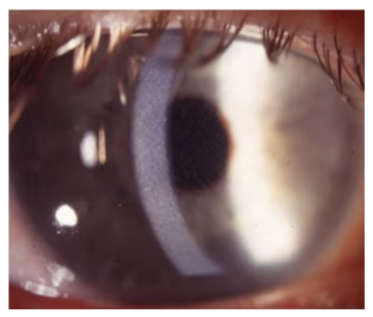
Image courtesy of William Trattler, MD.
图片由医学博士 William Trattler 提供。
Figure 2: Grade 3 diffuse lamellar keratitis
图 2:3 级弥漫性板层性角膜炎
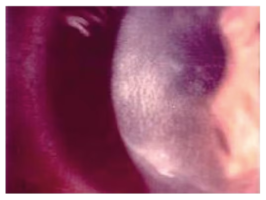
Image courtesy of William Trattler, MD.
图片由医学博士 William Trattler 提供。
Some studies have indicated a higher frequency of DLK in femtosecond LASIK cases compared to microkeratome8,27,45 with an incidence of Stage 1 or 2 DLK as high as 10.6% of patients, though all responded to steroids without visual consequence.27 New studies using updated femtosecond laser models suggest that the incidence is similar to LASIK performed with the microkeratome.46Patients are often asymptomatic but can present with pain or decreased vision. Factors that increase the risk of DLK are blood in the interface and flap epithelial defects.5 If either of these two findings are found, topical corticosteroid use should be increased. Early treatment is paramount with a focus on topical corticosteroids and occasionally oral steroids. In early stages, many surgeons increase topical prednisolone 1% to every hour and consider oral prednisone. If Stage 3 DLK is present, the flap is lifted, scraped, irrigated, and cultured, with possible application of steroids to the stromal bed. When this condition is recognized, it is important to follow closely to avoid stromal melt.4 Late-onset DLK can also happen and has been reported to occur as late as 17 years after LASIK.47
一些研究表明,与微型角膜刀相比 8,27,45 ,飞秒 LASIK 病例的 DLK 发生率更高,1 期或 2 期 DLK 的发生率高达 10.6%,尽管所有患者都对类固醇有反应,但没有视觉后果。 27 使用更新的飞秒激光模型的新研究表明,发生率与使用微角膜切术进行的LASIK相似。 46 患者通常无症状,但可表现为疼痛或视力下降。增加DLK风险的因素是界面出血和皮瓣上皮缺损。 5 如果发现这两种发现中的任何一种,应增加局部皮质类固醇的使用。早期治疗至关重要,重点是局部皮质类固醇,偶尔口服类固醇。在早期阶段,许多外科医生每小时增加 1% 的局部泼尼松龙,并考虑口服泼尼松龙。如果存在第 3 阶段 DLK,则提起、刮擦、冲洗和培养皮瓣,并可能在基质床上应用类固醇。当认识到这种情况时,重要的是要密切关注以避免基质熔化。 4 迟发性 DLK 也可能发生,据报道,迟至 LASIK 后 17 年才发生。 47
Infectious keratitis 感染性角膜炎
Infectious keratitis is a rare complication following LASIK but one of the most dreaded (see Figure 3). It is not specific to femtosecond laser. Studies have reported an incidence of 0–1.5%48 in LASIK patients and one retrospective case-control study looking at over 500,000 post LASIK patients found an incidence of 0.0046%.49 Presenting symptoms can occur acutely or over days to weeks and include decreased vision, erythema, photophobia, and pain. Bacterial keratitis occurs earlier, typically within 3–5 days, while atypical infections such as mycobacteria or fungal present after a few weeks. Treatment is typically with flap lift and irrigation, culture, broad-spectrum antibiotics, and possible flap amputation if necessary.50 Prevention includes aseptic technique, good lid hygiene, broad spectrum antibiotics in the early postoperative period, and continuous evaluation of sterile technique and instruments.
感染性角膜炎是LASIK术后一种罕见的并发症,但也是最可怕的并发症之一(见图3)。它不是飞秒激光器所特有的。研究表明,LASIK患者的发病率为0-1.5%, 48 一项针对超过500,000名LASIK术后患者的回顾性病例对照研究发现,发病率为0.0046%。 49 首发症状可急性发作,也可持续数天至数周,包括视力下降、红斑、畏光和疼痛。细菌性角膜炎发生得更早,通常在 3-5 天内发生,而分枝杆菌或真菌等非典型感染会在几周后出现。治疗通常包括皮瓣提升和冲洗、培养、广谱抗生素,必要时可能进行皮瓣截肢。 50 预防包括无菌技术、良好的盖子卫生、术后早期的广谱抗生素以及对无菌技术和器械的持续评估。
Figure 3: Infectious keratitis
图3:感染性角膜炎
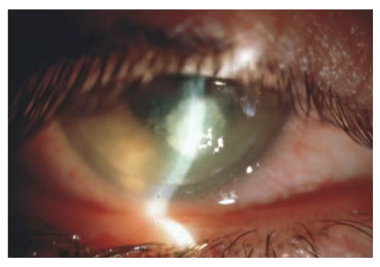
Image courtesy of William Trattler, MD.
图片由医学博士 William Trattler 提供。
Pressure-induced stromal keratitis
压力诱发的间质性角膜炎
PISK occurs as a result of increased intraocular pressure, typically with prolonged steroid use. In this condition, fluid can accumulate in the interface, leading to falsely low readings which can delay the diagnosis. Since the fluid amount is often small, it results in diffuse haziness in the interface and stroma, with no obvious fluid layer, though it can occasionally result in a visible fluid cleft separating the stromal cleavage plain.51,52 It has been reported to occur acutely in the postoperative setting and as a delayed complication, with some cases occurring years after the original LASIK surgery.53,54 As PISK is frequently misdiagnosed as DLK, further use of steroids can exacerbate the clinical condition. Routine IOP checks for patients on prolonged postoperative steroids is essential to diagnose this condition. However, interface fluid can lead to falsely low IOP, necessitating peripheral corneal pressure measurements and maintaining suspicion for the presence this condition. Treatment is typically to remove the steroid and consider a pressure-lowering drop as the IOP will follow suit, resolving the underlying problem.50
PISK的发生是由于眼压升高,通常是长期使用类固醇。在这种情况下,液体会积聚在界面中,导致读数过低,从而延迟诊断。由于液体量通常很小,因此导致界面和基质中弥漫性浑浊,没有明显的液体层,尽管偶尔会导致可见的液体裂隙将基质解理平原分开。 51,52 据报道,它在术后环境中急性发生,并作为延迟并发症发生,有些病例发生在最初的 LASIK 手术后数年。 53,54 由于 PISK 经常被误诊为 DLK,进一步使用类固醇会加剧临床状况。对长期术后类固醇患者进行常规眼压检查对于诊断这种疾病至关重要。然而,界面液可导致假性眼压低,需要测量外周角膜压并怀疑是否存在这种情况。治疗通常是去除类固醇并考虑降压,因为眼压也会随之下降,从而解决潜在的问题。 50
Central toxic keratopathy
中枢性中毒性角膜病变
Central toxic keratopathy is a rare non-inflammatory central corneal opacification that is acute and non-inflammatory in nature (see Figure 4). It occurs within days of stromal ablation procedures. The etiology is unknown but it presents acutely and does not worsen, unlike many of the other interface processes. It presents without pain, which can help distinguish it from DLK. Some surgeons attempt aggressive topical steroid use or flap irrigation, though interventions have not been shown to improve the final outcomes.50,55,56,46 The central opacity often spontaneously resolves in 2–18 months without intervention.
中枢性中毒性角膜病是一种罕见的非炎症性中央角膜混浊,性质为急性且非炎症性(见图4)。它发生在基质消融手术后的几天内。病因不明,但与许多其他界面过程不同,病因急性且不会恶化。它表现为无痛,这有助于将其与DLK区分开来。一些外科医生尝试积极局部使用类固醇或皮瓣冲洗,但干预措施尚未被证明可以改善最终结果。 50,55,56,46 中心性混浊通常在 2-18 个月内自行消退,无需干预。
Figure 4: Central toxic keratopathy
图 4:中枢性中毒性角膜病变

Discussion 讨论
The introduction of femtosecond laser technology has created a more reproducible and safe LASIK procedure and its use has increased over time. Microkeratomes have also improved with the rates of various complications, decreasing in both femtosecond and microkeratome-assisted LASIK cases. Over time, techniques, treatments and technologies have continued to improve, resulting in fewer complications.
飞秒激光技术的引入创造了一种更可重复和安全的LASIK手术,并且其使用随着时间的推移而增加。微角膜切口也随着各种并发症的发生率而改善,在飞秒和微角膜切术辅助LASIK病例中均有所下降。随着时间的流逝,技术、治疗方法和技术不断改进,导致并发症减少。
The next wave of refractive treatment, ReLEx/SMILE (small incision lenticule extraction; Carl Zeiss Meditec, Germany) has arrived and can only be accomplished with femtosecond technology. Due to this technology and past femtosecond trends, it is our belief that femtosecond laser use will continue to increase.
下一波屈光治疗,ReLEx/SMILE(小切口透镜拔除术;Carl Zeiss Meditec,德国)已经到来,只能通过飞秒技术来实现。由于这项技术和过去的飞秒趋势,我们相信飞秒激光的使用将继续增加。
Conclusions 结论
Femtosecond laser-assisted flap creation has improved the predictability and thickness of LASIK flaps making LASIK safer. Compared to microkeratome-created flaps, there are fewer complications overall, though femtosecond laser has resulted in a few new complications. It is important for any surgeon performing femtosecond laser-assisted LASIK to be aware of all possible intraoperative and postoperative complications. While the complications are applicable to all femtosecond lasers, it is essential that the surgeon also review the mechanism of suction and flap creation for the specific unit being used to carry out the procedure, as each model has different features that may help reduce certain complications but could predispose the patient to others. In addition to gaining a clear understanding of the mechanisms specific to the laser being used, by reviewing all potential procedural complications and ensuring they have the knowledge to manage any complications that may arise, surgeons can utilize femtosecond laser technology in a way that enables them to perform safe, reliable LASIK. ⬛
飞秒激光辅助皮瓣的创建提高了LASIK皮瓣的可预测性和厚度,使LASIK更安全。与微角膜膜瓣相比,尽管飞秒激光导致了一些新的并发症,但总体并发症较少。对于任何进行飞秒激光辅助LASIK手术的外科医生来说,了解所有可能的术中和术后并发症非常重要。虽然并发症适用于所有飞秒激光器,但外科医生还必须审查用于执行手术的特定单元的抽吸和皮瓣产生机制,因为每种型号都有不同的功能,可能有助于减少某些并发症,但可能使患者易患其他并发症。除了清楚地了解所使用的激光的特定机制外,通过审查所有潜在的手术并发症并确保他们具备管理可能出现的任何并发症的知识,外科医生还可以利用飞秒激光技术,使他们能够进行安全、可靠的 LASIK。⬛


