 Tianqi Lyu1
 Yuanbin Lin1
 Kerong Wu2Zhanglei Cao1Qian Zhang1Jianping Zheng1* 柳天琦 1 林元斌 1 吴克荣 2 曹章磊 1 张倩 1 郑剑平 1 *
- 1Cixi Institute of Biomedical Engineering, Ningbo Institute of Materials Technology and Engineering, Chinese Academy of Science (CAS), Ningbo, China
1 中国科学院宁波材料技术与工程研究所慈溪生物医学工程研究所,中国,宁波 - 2Department of Urology, Ningbo First Hospital, School of Medicine Ningbo University, Zhejiang University Ningbo Hospital, Ningbo, China
2 宁波大学医学院附属宁波第一医院泌尿外科,浙江大学附属宁波医院,宁波,中国
Bladder cancer is among the most common malignant tumors with highly heterogeneous molecular characteristics. Despite advancements of the available therapeutic options, several bladder cancer patients exhibit unsatisfactory clinical outcomes. The lack of specific biomarkers for effective targeted therapy or immunotherapy remains a major obstacle in treating bladder cancer. The rapid development of single-cell techniques is transforming our understanding of the intra-tumoral heterogeneity, thereby providing us with a powerful high-throughput sequencing tool that can reveal tumorigenesis, progression, and invasion in bladder tumors. In this review, we summarise and discuss how single-cell sequencing technologies have been applied in bladder cancer research, to advance our collective knowledge on the heterogeneity of bladder tumor cells, as well as to provide new insights into the complex ecosystem of the tumor microenvironment. The application of single-cell approaches also uncovers the therapeutic resistance mechanism in bladder cancer and facilitates the detection of urinary-exfoliated tumor cells. Moreover, benefiting from the powerful technical advantages of single-cell techniques, several key therapeutic targets and prognostic models of bladder cancer have been identified. It is hoped that this paper can provide novel insights into the precision medicine of bladder cancer.
膀胱癌是最常见的恶性肿瘤之一,具有高度异质性的分子特征。尽管现有的治疗方案不断进步,但仍有一些膀胱癌患者的临床治疗效果不尽人意。缺乏有效靶向治疗或免疫疗法的特异性生物标志物仍然是治疗膀胱癌的主要障碍。单细胞技术的快速发展改变了我们对肿瘤内部异质性的认识,从而为我们提供了一种强大的高通量测序工具,可以揭示膀胱肿瘤的发生、发展和侵袭过程。在这篇综述中,我们总结并讨论了单细胞测序技术在膀胱癌研究中的应用情况,这些应用增进了我们对膀胱肿瘤细胞异质性的集体认识,并为我们了解肿瘤微环境的复杂生态系统提供了新的视角。单细胞方法的应用还揭示了膀胱癌的治疗耐药机制,并有助于检测尿道脱落的肿瘤细胞。此外,得益于单细胞技术强大的技术优势,一些膀胱癌的关键治疗靶点和预后模型已被确定。希望本文能为膀胱癌的精准医疗提供新的见解。
Introduction 导言
Bladder cancer is the most common malignant tumor that affects the urinary system and is associated with high morbidity and mortality (Antoni et al., 2017). Several risk factors such as sex, age, lifestyle, pathogenic microbial infections, and drugs may augment the bladder cancer risk (Lin et al., 2006; Letašiová et al., 2012; Burger et al., 2013; Berdik, 2017). Despite advancements of the available therapeutic approaches, the clinical outcomes of bladder cancer patients remain unsatisfactory, and the corresponding 5-year survival rate remains almost unchanged. Bladder cancer is one of the highly heterogeneous human cancers, often involving frequent genomic alterations and molecular subtype diversity, and it may be the first cause of therapeutic failure; therefore, the elucidation of the molecular and cellular heterogeneity of bladder cancer is necessary (Genitsch et al., 2019). Probably, an in-depth understanding of the unique molecular characteristics and cellular heterogeneity involved in the initiation, development, and pathogenesis of bladder cancer can improve the clinical treatment and prognosis, as well as contribute to the promotion of its diagnosis and treatment towards precision medicine.
膀胱癌是影响泌尿系统的最常见恶性肿瘤,与高发病率和高死亡率相关(Antoni 等人,2017 年)。性别、年龄、生活方式、病原微生物感染和药物等多种风险因素都可能增加膀胱癌风险(Lin 等人,2006 年;Letašiová 等人,2012 年;Burger 等人,2013 年;Berdik,2017 年)。尽管现有的治疗方法取得了进步,但膀胱癌患者的临床治疗效果仍不令人满意,相应的 5 年生存率几乎没有变化。膀胱癌是高度异质性的人类癌症之一,通常涉及频繁的基因组改变和分子亚型多样性,它可能是治疗失败的首要原因;因此,阐明膀胱癌的分子和细胞异质性是必要的(Genitsch 等人,2019 年)。深入了解膀胱癌发病、发展和致病过程中的独特分子特征和细胞异质性,可能会改善临床治疗和预后,并有助于推动膀胱癌的诊断和治疗向精准医学方向发展。
Over the past few decades, with the rapid development of next-generation sequencing and molecular biology technologies, research on the biological characteristics and genotyping of bladder cancer has made considerable strides (Pietzak et al., 2017; Roy et al., 2017; Wu et al., 2019; Li et al., 2021a; Tran et al., 2021). Bladder cancer can be categorised into three subtypes based on its histological features: urothelial carcinoma, squamous cell carcinoma, and adenocarcinoma. Of these subtypes, urothelial tumors account for approximately 90% of cases, while squamous and glandular-type tumors are rare (Kantor et al., 1988; Knowles and Hurst, 2015). The 2016 World Health Organisation grading system has stratified bladder cancer into non-muscle-invasive bladder cancer (NMIBC) and muscle-invasive bladder cancer (MIBC) (Compérat et al., 2019). At the first clinical diagnosis, approximately 85% of bladder cancer patients are diagnosed with NMIBC, and up to 80% of these patients have at least one recurrence, whereas disease progression into MIBC occurs in approximately 30% of these patients (Batista et al., 2020; van den Bosch and Alfred Witjes, 2011). It is difficult for doctors to predict the risk of recurrence and progression of NMIBC based on the conventional classification system.
过去几十年来,随着新一代测序和分子生物学技术的快速发展,膀胱癌的生物学特征和基因分型研究取得了长足的进步(Pietzak 等,2017;Roy 等,2017;Wu 等,2019;Li 等,2021a;Tran 等,2021)。根据组织学特征,膀胱癌可分为三个亚型:尿路上皮癌、鳞状细胞癌和腺癌。在这些亚型中,尿路上皮癌约占 90%,而鳞癌和腺癌则很少见(Kantor 等人,1988 年;Knowles 和 Hurst,2015 年)。2016 年世界卫生组织分级系统将膀胱癌分为非肌层浸润性膀胱癌(NMIBC)和肌层浸润性膀胱癌(MIBC)(Compérat 等人,2019 年)。在首次临床诊断时,约有85%的膀胱癌患者被诊断为NMIBC,其中多达80%的患者至少有一次复发,而这些患者中约有30%的人病情恶化为MIBC(Batista等人,2020年;van den Bosch和Alfred Witjes,2011年)。医生很难根据传统的分类系统预测 NMIBC 的复发和进展风险。
As of today, enormous efforts are being undertaken to outline the diverse molecular subtypes and collect crucial biological information on bladder cancer. Several researchers have proposed that the molecular and genetic heterogeneities of bladder cancer cells are critical to diagnosis and treatment of the disease (Thomsen et al., 2017; Audenet et al., 2018; Sjödahl et al., 2019; Minoli et al., 2020). However, despite these advances, clinical therapies based on the molecular subtypes and genetic features of bladder cancer often show several limitations such as poor efficacy, drug resistance, and recurrence due to tumor heterogeneity (Casadei et al., 2019; Kamoun et al., 2019; Zhu et al., 2020). In general, the molecular mechanisms for the initiation and development of bladder cancer remain elusive, and the current molecular subtyping systems do not depict the cellular heterogeneity of bladder carcinoma. In the era of precision medicine, we should consider the vital role played by inter-tumoral and intra-tumoral heterogeneities. Decoding the heterogeneity of bladder cancer cells at the genetic and cellular levels, refinement of the molecular types, and discovering new ways to overcome resistance are the main issues that should be addressed in the studies on bladder cancer.
时至今日,人们正付出巨大努力,勾勒出膀胱癌的各种分子亚型,并收集有关膀胱癌的重要生物学信息。一些研究人员提出,膀胱癌细胞的分子和遗传异质性对于疾病的诊断和治疗至关重要(Thomsen 等人,2017 年;Audenet 等人,2018 年;Sjödahl 等人,2019 年;Minoli 等人,2020 年)。然而,尽管取得了这些进展,基于膀胱癌分子亚型和遗传特征的临床疗法往往表现出一些局限性,如疗效不佳、耐药性以及肿瘤异质性导致的复发等 ( Casadei 等人,2019;Kamoun 等人,2019;Zhu 等人,2020)。总体而言,膀胱癌发生和发展的分子机制仍然难以捉摸,目前的分子亚型系统也无法描述膀胱癌的细胞异质性。在精准医疗时代,我们应该考虑肿瘤间和肿瘤内异质性的重要作用。从基因和细胞层面解码膀胱癌细胞的异质性,细化分子类型,发现克服耐药性的新方法,是膀胱癌研究应解决的主要问题。
In 2009, Tang et al. reported the single-cell RNA sequencing (scRNA-seq) technology for the first time, which helped solve the problem of cell heterogeneity that was difficult to be solved by bulk RNA sequencing (Tang et al., 2009). The rapid developments in single-cell techniques allow us to interrogate the transcriptional, genomic, proteomic, epigenomic, metabolic, and multi-omic characteristics of thousands of individual cells (Kashima et al., 2020) (Table 1). This powerful high-resolution and high-throughput sequencing tool has revolutionised our understanding of cancer biology and tumor cytology (Stuart and Satija, 2019; Xing et al., 2020). These technologies have helped in successfully deciphering the compositional complexity and clonal heterogeneity in tumors (Lei et al., 2021). Indeed, single-cell techniques have multiple applications in cancer research, which include the identification of dynamic gene expression profiles during tumor progression, discovery of novel cell subpopulations, determination of cell status and phenotype switches, analysis of the regulatory pathways of key genes, and identification of potential therapeutic targets (Papalexi and Satija, 2018) (Figure 1). Owing to the excellent technical advantages, single-cell techniques have undoubtedly promoted the precision and accuracy of molecular cancer research to new levels. The past decade has seen unprecedented advances in bladder cancer studies, which can be ascribed mainly to single-cell techniques (Table 2). In this review, we summarise pioneering research in the field and discuss the latest progress in the aspects such as cellular heterogeneity, tumor microenvironment (TME), drug resistance, and prognosis of bladder cancer. All of these further inspire and foster research on clinically relevant diagnosis, prognosis prediction, targeted therapy, and personalised therapeutic strategies for bladder cancer.
2009 年,Tang 等人首次报道了单细胞 RNA 测序(scRNA-seq)技术,该技术有助于解决大量 RNA 测序难以解决的细胞异质性问题(Tang 等人,2009 年)。单细胞技术的飞速发展使我们能够研究成千上万个单细胞的转录、基因组、蛋白质组、表观遗传组、代谢和多组学特征(Kashima 等人,2020 年)(表 1)。这种强大的高分辨率和高通量测序工具彻底改变了我们对癌症生物学和肿瘤细胞学的认识(Stuart 和 Satija,2019 年;Xing 等人,2020 年)。这些技术有助于成功破译肿瘤的组成复杂性和克隆异质性(Lei 等,2021 年)。事实上,单细胞技术在癌症研究中有多种应用,其中包括鉴定肿瘤进展过程中的动态基因表达谱、发现新型细胞亚群、确定细胞状态和表型转换、分析关键基因的调控通路以及鉴定潜在的治疗靶点(Papalexi 和 Satija,2018 年)(图 1)。凭借出色的技术优势,单细胞技术无疑将癌症分子研究的精度和准确性提升到了新的水平。过去十年,膀胱癌研究取得了前所未有的进展,这主要归功于单细胞技术(表 2)。在这篇综述中,我们总结了该领域的开创性研究,并讨论了膀胱癌细胞异质性、肿瘤微环境(TME)、耐药性和预后等方面的最新进展。 所有这些都进一步激发和促进了有关膀胱癌临床诊断、预后预测、靶向治疗和个性化治疗策略的研究。

TABLE 1. Single-cell omics technologies.
表 1.单细胞 omics 技术。
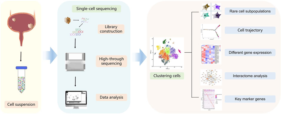
FIGURE 1. Schematic diagram of the single-cell sequencing workflow. Cells are dissolved from bladder tumors into single-cell suspension. Single cells are then processed into the reaction system followed by reverse transcription, amplification, library construction and sequencing. Sequencing data are processed via bioinformatic analysis such as cell clustering, cell trajectory, different gene expression, interactome or key marker genes analysis.
图 1.单细胞测序工作流程示意图。将膀胱肿瘤中的细胞溶解成单细胞悬浮液。然后将单细胞放入反应系统,进行反转录、扩增、文库构建和测序。测序数据经生物信息分析处理,如细胞聚类、细胞轨迹、不同基因表达、相互作用组或关键标记基因分析。
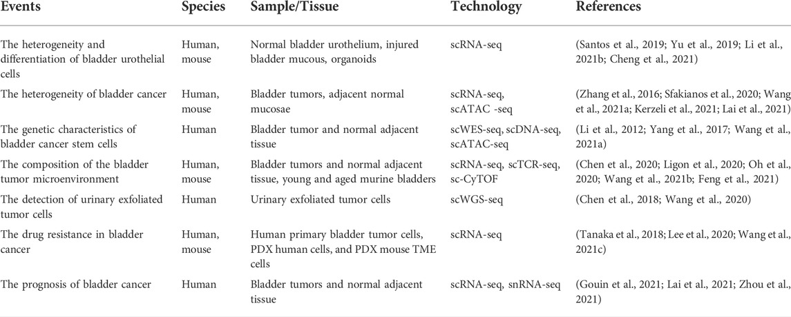
TABLE 2. Summary of the studies in bladder cancer using single-cell technologies.
表 2.使用单细胞技术的膀胱癌研究摘要。
Uncovering the heterogeneity of bladder urothelial cells through single-cell sequencing
通过单细胞测序揭示膀胱尿路上皮细胞的异质性
The urothelium of the urinary bladder is a particular hierarchically organised tissue that serves as the strongest urine–blood barrier in the body. The adult urothelium can be histologically classified as the transitional epithelium consisting of three types of cells, namely basal, intermediate, and umbrella cells (Figure 2). Among these cells, the basal cells are small and polygonal and form a single layer that directly contacts the basement membrane. Intermediate cells are pyriform and form multiple cell layers. Umbrella cells are large binuclear or multinuclear cubic cells that form a single layer in direct contact with the urinary space (Apodaca, 2004; Khandelwal et al., 2009; Ho et al., 2012). Usually, the bladder urothelium regenerates rapidly upon injury, which depends on the proliferation of urothelial stem cells (Ho et al., 2012). Accumulating evidence suggests that minor subpopulations of basal cells, characterised by the expressions of keratin 5 (KRT5) and keratin 14 (KRT14), possess self-renewal capacity and give rise to all cell types of the urothelium during natural and injury-induced regeneration processes, which represents the cellular origins of urothelial carcinoma (Shin et al., 2011; Papafotiou et al., 2016). Reported research demonstrates that a vast majority of tumor lesions of the bladder arise from the urothelial cells (Castillo-Martin et al., 2010). Therefore, elucidation of the primitive cellular differentiation status, stem cell differentiation, and genetic alteration characteristics of bladder urothelial may provide critical biological insights for understanding the molecular mechanism of associated bladder diseases.
膀胱的尿路上皮是一种特殊的分层组织,是人体最坚固的尿液-血液屏障。成人尿路上皮在组织学上可分为过渡上皮,由基底细胞、中间细胞和伞状细胞三种类型的细胞组成(图 2)。在这些细胞中,基底细胞体积小,呈多角形,形成单层,直接与基底膜接触。中间细胞呈梨形,形成多个细胞层。伞细胞是大的双核或多核立方细胞,形成单层,直接与泌尿空间接触(Apodaca,2004;Khandelwal 等人,2009;Ho 等人,2012)。通常,膀胱尿路上皮在受伤后会迅速再生,这取决于尿路上皮干细胞的增殖(Ho 等人,2012 年)。越来越多的证据表明,以角蛋白5(KRT5)和角蛋白14(KRT14)的表达为特征的基底细胞小亚群具有自我更新能力,并在自然和损伤诱导的再生过程中产生尿路上皮的所有细胞类型,这代表了尿路上皮癌的细胞起源(Shin等人,2011年;Papafotiou等人,2016年)。研究报告表明,绝大多数膀胱肿瘤病变来源于尿路上皮细胞(Castillo-Martin 等人,2010 年)。因此,阐明膀胱尿路上皮细胞的原始细胞分化状态、干细胞分化和基因改变特征可为了解相关膀胱疾病的分子机制提供重要的生物学见解。
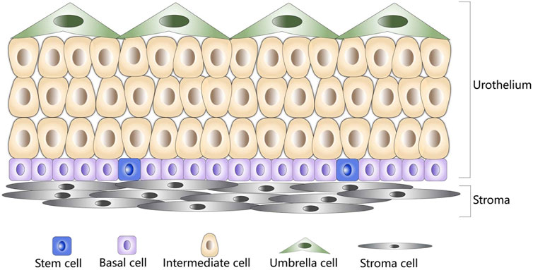
FIGURE 2. Schematic diagram of normal bladder urothelium. Stem cells are shown in blue, basal cells are shown in purple, intermediate cells are shown in brown, umbrella cells are shown in green, and stroma cells are shown in gray.
图 2.正常膀胱尿路上皮细胞示意图。干细胞为蓝色,基底细胞为紫色,中间细胞为棕色,伞状细胞为绿色,基质细胞为灰色。
To gain an insight into the cell subtypes and urothelial differentiation characteristics, Li et al. performed scRNA-seq to detect urothelial cells digested and isolated from the mouse bladder urothelium (Li et al., 2021b). The authors discovered that the ASPM-labelled basal-like cells might be the progenitor cells of the other subpopulations that could participate in regeneration of the bladder urothelium. In particular, a novel superficial-like cell population (Plxna4+ urothelial cells) may play a priming role in the initiation of resisting inflammation and repairing of the responses of the bladder urothelium. Yu et al. compared the single-cell transcriptomic maps of human and mouse bladders to determine both the conservative and heterogeneous aspects of human and mouse bladder evolution (Yu et al., 2019). The authors identified two new types of human bladder cells: ADRA2A+ and HRH2+ interstitial cells and TNNT1+ epithelial cells. Of these, ADRA2A+ interstitial cells appear to play a special role in the human bladder, while TNNT1+ epithelial cells are also present in rat and mouse bladder tissues. Developmental trajectory analysis revealed that the bladder epithelial cells could be transformed from basal cells to intermediate cells and then into umbrella cells or TNNT1+ epithelial cells. Another study recapitulated the differentiation and function of urothelial cells by establishing mouse urothelial organoids, wherein the authors identified five cell clusters by utilising scRNA-seq and reported that the CD49f/CD44 high-labelled urothelial cell subpopulation exhibits the highest organoid-forming potential (Santos et al., 2019; Yu et al., 2019). In previous studies, CD49f and CD44 have been proposed as markers for aggressive basal/squamous bladder cancer subtypes (Lerner et al., 2016; Sjödahl et al., 2017). Notably, the investigation unveiled the essential role of Notch signalling in the differentiation of normal urothelial cells, which was elusive in the past bulk organoid transcriptomics studies. Furthermore, the study confirmed that gene mutations in the Notch pathway could induce urothelial tumors.
为了深入了解细胞亚型和尿路上皮的分化特征,Li 等人采用 scRNA-seq 技术检测了从小鼠膀胱尿路上皮中消化和分离出来的尿路上皮细胞(Li 等人,2021b)。作者发现,ASPM 标记的基底样细胞可能是其他亚群的祖细胞,可参与膀胱尿路上皮的再生。特别是,一个新的表层样细胞群(Plxna4 + 尿路上皮细胞)可能在膀胱尿路上皮开始抵抗炎症和修复反应的过程中发挥了先导作用。Yu等人比较了人类和小鼠膀胱的单细胞转录组图谱,以确定人类和小鼠膀胱进化的保守性和异质性方面(Yu等人,2019年)。作者发现了两种新型人类膀胱细胞:ADRA2A + 和 HRH2 + 间质细胞以及 TNNT1 + 上皮细胞。其中,ADRA2A + 间质细胞似乎在人类膀胱中发挥着特殊作用,而TNNT1 + 上皮细胞也存在于大鼠和小鼠的膀胱组织中。发育轨迹分析表明,膀胱上皮细胞可从基底细胞转变为中间细胞,然后转变为伞状细胞或 TNNT1 + 上皮细胞。另一项研究通过建立小鼠尿路上皮器官组织重现了尿路上皮细胞的分化和功能,作者利用scRNA-seq鉴定了五个细胞群,并报告说CD49f/CD44高标记的尿路上皮细胞亚群表现出最高的器官组织形成潜能(Santos等人,2019;Yu等人,2019)。 在以前的研究中,CD49f和CD44被认为是侵袭性基底/鳞状膀胱癌亚型的标志物(Lerner等人,2016年;Sjödahl等人,2017年)。值得注意的是,该研究揭示了Notch信号在正常尿路上皮细胞分化中的重要作用,而这在过去的大块类器官转录组学研究中是难以捉摸的。此外,该研究还证实,Notch通路中的基因突变可诱发尿道肿瘤。
Recently, Cheng et al. explored the cell heterogeneity involved in the repair and regeneration processes of bladder urothelial cells and focused on the crosstalk between cell subsets (Cheng et al., 2021). When compared with the normal urothelial cells, two cycling cell populations (i.e., basal cells and luminal cells) were highly enriched under injured conditions. A study demonstrated that urothelium regeneration is mediated by distinct division patterns of the cycling basal and intermediate cells (Wang et al., 2018); in this study, the single-cell analysis confirmed that this discrepancy of cell cycle progression might be attributable to the difference in cell adhesion (Cheng et al., 2021). By analysing the lineage relationship among urothelial cell subpopulations, the authors found that cycling basal cells and cycling intermediate cells represent two different states of the urothelium. Notably, cycling intermediate cells could act as the direct progenitors of superficial cells, where the latter was directly repaired by the former, which verifies the previous hypothesis. According to cell–cell communication analyses, Acta2+Cd34+ myofibroblasts have the most significant influence on the proliferation of urothelial cells, and both Bmp and Fgfr signaling are involved in this process (Cheng et al., 2021). These results together supported the view that epithelial–myofibroblast crosstalk plays a key role in urothelial homeostasis and repair.
最近,Cheng 等人探索了膀胱尿路上皮细胞修复和再生过程中的细胞异质性,并重点研究了细胞亚群之间的串扰(Cheng 等人,2021 年)。与正常尿路上皮细胞相比,两种循环细胞群(即基底细胞和管腔细胞)在损伤条件下高度富集。一项研究表明,尿路上皮的再生是由循环基底细胞和中间细胞不同的分裂模式介导的(Wang等人,2018年);在这项研究中,单细胞分析证实了细胞周期进展的这种差异可能是由于细胞粘附性的不同造成的(Cheng等人,2021年)。通过分析尿路上皮细胞亚群之间的系谱关系,作者发现循环基底细胞和循环中间细胞代表了尿路上皮的两种不同状态。值得注意的是,循环中间细胞可以作为表层细胞的直接祖细胞,后者直接由前者修复,这验证了之前的假设。根据细胞间通讯分析,Acta2 + Cd34 + 肌成纤维细胞对尿路上皮细胞的增殖影响最大,Bmp 和 Fgfr 信号都参与了这一过程(Cheng 等人,2021 年)。这些结果共同支持了上皮细胞-肌成纤维细胞串联在尿道稳态和修复中发挥关键作用的观点。
In summary, these results further reinforce the conclusion that bladder urothelial cells are highly heterogeneous and provide fundamental insights into the correlation between diverse cell phenotypes of the bladder urothelium and bladder diseases.
总之,这些结果进一步证实了膀胱尿路上皮细胞具有高度异质性的结论,并为了解膀胱尿路上皮细胞的不同细胞表型与膀胱疾病之间的相关性提供了基本见解。
Single-cell sequencing in the cellular heterogeneity of bladder cancer
单细胞测序在膀胱癌细胞异质性研究中的应用
Discerning the heterogeneity of individual tumor cells carries clinical implications for predicting prognosis and treatment responses of bladder cancer. For instance, in the murine and human bladder cancer model systems, single-cell transcriptome analyses revealed the presence of multi-lineage gene expression phenotypes. In addition, among the epithelial cell clusters, cell subpopulations with concomitantly pronounced expressions of basal, luminal, and EMT claudin-related genes were characterised, and the cellular expression of these lineage markers was found to be dynamic either during tumor progression or in response to the treatment. Analysis of transplantable MIBC models demonstrated that these specific multi-lineage subtypes could define the cancer cells capable of tumor formation and lineage plasticity, which could be distinguished by the surface antigen CD49f in conjunction with the epithelial markers (Sfakianos et al., 2020). Finally, investigators affirmed the exact composition of the intra-tumoral cells derived from multi-lineage subpopulations as showing inherent heterogeneity and lineage plasticity of the epithelium. Data from a recent study revealed the heterogeneity of epithelium in bladder cancer patients with different pathological tumor stages (Lai et al., 2021). According to previous reports, bladder cancer is a urinary tract epithelium-derived cancer, and epithelial–mesenchymal transition (EMT) promotes the transition from NMIBC to MIBC (Cao et al., 2020). The malignant cell-specific gene expression suggested that basal-like cells were invasive precursors that displayed EMT characteristics in NMIBC. Recently, Wang et al. elucidated the spectrum of EMT associated with bladder cancer subtypes and identified that TCF7 promotes EMT in corroboration with single-cell ATAC sequencing (scATAC-seq) (Wang et al., 2021a). This evidence suggested that the epithelial cells represent the malignant cells of bladder cancer and that the upregulation of EMT is essential for driving local bladder tumor cells’ invasion and progression.
辨别单个肿瘤细胞的异质性对预测膀胱癌的预后和治疗反应具有临床意义。例如,在小鼠和人类膀胱癌模型系统中,单细胞转录组分析显示存在多系基因表达表型。此外,在上皮细胞簇中,基底、管腔和 EMT claudin 相关基因同时明显表达的细胞亚群也得到了表征,而且发现在肿瘤进展过程中或对治疗做出反应时,这些系标志物的细胞表达是动态的。对可移植的 MIBC 模型的分析表明,这些特定的多系亚型可以确定能够形成肿瘤和具有系可塑性的癌细胞,这些癌细胞可以通过表面抗原 CD49f 与上皮标记物结合来区分(Sfakianos 等人,2020 年)。最后,研究人员肯定了源自多系亚群的瘤内细胞的确切组成,这显示了上皮细胞固有的异质性和系可塑性。最近的一项研究数据显示,不同病理阶段的膀胱癌患者上皮细胞具有异质性(Lai 等人,2021 年)。根据之前的报道,膀胱癌是一种尿路上皮源性癌症,上皮-间质转化(EMT)促进了从NMIBC到MIBC的转变(Cao等人,2020)。恶性细胞特异性基因表达表明,基底样细胞是NMIBC中显示EMT特征的侵袭性前体细胞。最近,Wang 等人发现,在 NMIBC 中,基底样细胞是具有 EMT 特征的侵袭性前体细胞。 王等人,2021a)阐明了与膀胱癌亚型相关的EMT谱,并通过单细胞ATAC测序(scATAC-seq)确定了TCF7促进EMT。这些证据表明,上皮细胞代表了膀胱癌的恶性细胞,而 EMT 的上调对于推动局部膀胱肿瘤细胞的侵袭和进展至关重要。
To further investigate the tumor heterogeneity involved in the transition from NMIBC to MIBC, Kerzeli et al. established a novel urothelial carcinoma model of Hgf-Cdk4R24C mice and identified eight heterogeneous cell clusters of urinary epithelium origin by utilising single-cell transcriptomic analyses (Kerzeli et al., 2021). Unlike the urothelial cells in healthy bladders, there were urothelial cell clusters with highly proliferative or cancer stemness gene expression characteristics in carcinogens-induced bladder tumors. Notably, these cells displaying cell cycle dysregulation gene expression profiles were considered asaggressive subclones. These results are in line with the reports of a study that observed intra-tumoral heterogeneity of human urothelial bladder cancer, identifying it as clonal mutations according to their genetic characteristics (Heide et al., 2019). Since the histopathological feature of Hgf-Cdk4R24C bladder tumors was urothelial carcinoma with squamous differentiation (UC/SCC), the proposed model would be a helpful tool for determining the tumor biological characteristics and the treatment response of UC/SCC.
为了进一步研究从NMIBC向MIBC转变过程中涉及的肿瘤异质性,Kerzeli等人建立了一种新型的Hgf-Cdk4R24C小鼠尿路上皮癌模型,并利用单细胞转录组分析鉴定了8个来源于尿路上皮的异质性细胞簇(Kerzeli等人,2021年)。与健康膀胱中的尿路上皮细胞不同,在致癌物质诱发的膀胱肿瘤中,尿路上皮细胞簇具有高度增殖或癌症干基因表达特征。值得注意的是,这些显示细胞周期失调基因表达谱的细胞被认为是侵袭性亚克隆。这些结果与一项观察到人类尿道膀胱癌瘤内异质性的研究报告一致,根据其遗传特征将其确定为克隆突变(Heide等人,2019年)。由于Hgf-Cdk4R24C膀胱肿瘤的组织病理学特征是具有鳞状分化的尿路上皮癌(UC/SCC),因此所提出的模型将有助于确定UC/SCC的肿瘤生物学特征和治疗反应。
Histologically, different from UC/SCC, tumors in bladder cancer patients with pure squamous cell components are defined as squamous cell carcinoma of the bladder (SCCB). Although SCCB accounts for <10% of primary bladder cancer, it shows frequent recurrence and metastasis when compared with urothelial carcinoma, which highlights the more complex intra-tumoral heterogeneity of SCCB (Minato et al., 2018; Lin et al., 2019). In 2016, a study conducted scRNA-seq on the tumor-normal paired tissues from an SCCB patient and revealed intra-tumoral heterogeneity and the potential mechanisms of SCCB (Zhang et al., 2016). The authors considered that genes with different expression patterns particularly enriched in the MAPK signaling pathway were involved in tumor evolution and heterogeneity formation. Especially, a keratinocyte-specific POU transcription factor, POU2F3, was associated with squamous epithelial stratification, indicating that POU2F3 may be a crucial biomarker of SCCB.
在组织学上,与尿路上皮癌/膀胱癌不同,膀胱癌患者的肿瘤中含有纯鳞状细胞成分,被定义为膀胱鳞状细胞癌(SCCB)。虽然SCCB在原发性膀胱癌中的占比小于10%,但与尿路上皮癌相比,SCCB却显示出频繁的复发和转移,这凸显了SCCB更复杂的瘤内异质性(Minato等,2018;Lin等,2019)。2016 年,一项研究对一名 SCCB 患者的肿瘤-正常配对组织进行了 scRNA-seq,揭示了 SCCB 的瘤内异质性和潜在机制 ( Zhang et al., 2016)。作者认为,具有不同表达模式的基因,尤其是富含 MAPK 信号通路的基因,参与了肿瘤的演变和异质性的形成。尤其是角朊细胞特异性 POU 转录因子 POU2F3 与鳞状上皮分层相关,表明 POU2F3 可能是 SCCB 的关键生物标志物。
In general, these findings reveal the complex intra-tumoral and inter-tumor heterogeneities of bladder cancer, which can explain its diverse molecular and clinical phenotypes. These identified aggressive subclones and biomarkers may help enhance our understanding of the progression of bladder cancer, thereby facilitating individualised diagnosis and treatment.
总之,这些发现揭示了膀胱癌复杂的瘤内和瘤间异质性,这可以解释其多样的分子和临床表型。这些已发现的侵袭性亚克隆和生物标志物可能有助于加深我们对膀胱癌进展的了解,从而促进个体化诊断和治疗。
Single-cell sequencing for investigating the genetic characteristics of bladder CSCs
利用单细胞测序研究膀胱造血干细胞的遗传特征
Cancer stem cells (CSCs) possess the capability of self-renewal and are responsible for the initiation and development of tumors. Past studies have suggested that bladder CSCs possess high tumorigenicity, drug resistance, metastasis, and typical biomarkers (Ho et al., 2012). As early as 2012, Li et al. employed the single-cell exome sequencing technology to analyse the genetic landscape of bladder carcinoma and deciphered the mechanism of its occurrence and development (Li et al., 2012). Their study suggested that bladder cancer cells are derived from a single ancestral cell that formed two distinct tumor cell subgroups in the subsequent evolutionary process. Furthermore, they demonstrated that the ancestral bladder cancer cells had a monoclonal phenotype with multiple mutation driver gene candidates, among which ATM, COL6A3, and KIAA1958 were discovered as novel subclone-specific genes. These candidate cancer-related genes could drive the initiation of carcinogenesis and the development of subsequent cell lineages involved in cancer progression, which best matches the clonal evolution model. These findings explained that the genesis of bladder cancer was multi-gene mutation drive and multi-factorial, which in turn provided evidence for deciphering how tumors evolve into difficult-to-treat metastases. However, there remains an ambiguity related to whether ancestral cells inferred by the authors represent the initial tumor cells (Heng et al., 2006; Li et al., 2012; Ren et al., 2018; Zhang and Zhang, 2020).
癌症干细胞(CSCs)具有自我更新能力,是肿瘤发生和发展的元凶。过去的研究表明,膀胱癌干细胞具有高致瘤性、耐药性、转移性和典型的生物标志物(Ho 等,2012)。早在2012年,Li等人就利用单细胞外显子组测序技术分析了膀胱癌的基因图谱,并破译了其发生和发展的机制(Li等人,2012)。他们的研究表明,膀胱癌细胞来源于一个祖先细胞,在随后的进化过程中形成了两个不同的肿瘤细胞亚群。此外,他们还证明祖先膀胱癌细胞具有单克隆表型,并有多个突变驱动基因候选,其中发现了 ATM、COL6A3 和 KIAA1958 等新型亚克隆特异性基因。这些与癌症相关的候选基因可驱动癌变的发生,以及参与癌症进展的后续细胞系的发展,这与克隆进化模型最为吻合。这些发现解释了膀胱癌的发生是由多基因突变驱动和多因素造成的,从而为破译肿瘤如何演变为难以治疗的转移瘤提供了证据。然而,作者们推断的祖先细胞是否代表最初的肿瘤细胞仍存在模糊之处(Heng等人,2006年;Li等人,2012年;Ren等人,2018年;Zhang和Zhang,2020年)。
To further investigate the genetic basis and origin of bladder CSCs, Yang et al. conducted single-cell sequencing on 59 cells from three human bladder cancer specimens and examined their phylogenetic status (Yang et al., 2017). Their research demonstrated that bladder CSCs originated from bladder epithelial stem cells or bladder cancer non-stem cells, thus providing genetic evidence to support the hypothesis of CSC origin. Moreover, 21 key gene mutations in bladder CSCs were discovered, which involved the genes related to cell cycle regulation, transcription regulation, chromatin remodeling, cell differentiation, and self-renewal. These specific mutations are critical to the acquisition of bladder CSC stemness. Moreover, the alterations of the three genes ARID1A, GPRC5A, and MLL2 play a crucial role in conferring stemness to the bladder cancer non-stem cells, as confirmed by co-mutation gene assays. Wang and others performed single-cell ATAC sequencing and found that bladder CSCs were enriched during tumor recurrence with elevated expression of EZH2. The specific molecular mechanism is that EZH2 maintains H3K27me3-mediated repression of the NCAM1 gene, thereby inactivating cellular invasiveness and stemness transcriptional programs (Wang et al., 2021a).
为了进一步研究膀胱干细胞的遗传基础和起源,Yang等人对来自三个人类膀胱癌标本的59个细胞进行了单细胞测序,并研究了它们的系统发育状况(Yang等人,2017)。他们的研究表明,膀胱干细胞起源于膀胱上皮干细胞或膀胱癌非干细胞,从而为支持干细胞起源假说提供了遗传学证据。此外,研究还发现了膀胱干细胞中的21个关键基因突变,涉及细胞周期调控、转录调控、染色质重塑、细胞分化和自我更新等相关基因。这些特定突变对膀胱干细胞干性的获得至关重要。此外,通过共突变基因检测证实,ARID1A、GPRC5A和MLL2这三个基因的改变在赋予膀胱癌非干细胞干性方面起着至关重要的作用。Wang等人进行了单细胞ATAC测序,发现膀胱癌干细胞在肿瘤复发过程中富集,EZH2表达升高。具体的分子机制是EZH2维持H3K27me3介导的对NCAM1基因的抑制,从而使细胞侵袭性和干性转录程序失活(Wang等人,2021a)。
Overall, several potential key genes related to the characteristics of bladder CSCs have been uncovered so far. Nevertheless, further studies on the function and clinical applications are warranted to determine whether these candidate genes can act as bladder cancer biomarkers to guide targeted therapy.
总之,迄今为止已经发现了几个与膀胱干细胞特征相关的潜在关键基因。然而,要确定这些候选基因能否作为膀胱癌生物标志物指导靶向治疗,还需要对其功能和临床应用进行进一步研究。
Single-cell sequencing reveals the complexity of the bladder TME
单细胞测序揭示膀胱TME的复杂性
The tumorigenesis, progression, and invasion of cancer cells are closely related to their surrounding microenvironment (Bryan, 2015; Song et al., 2019). Notably, bladder cancer is one of the least immune invasive cancers (Chen et al., 2017), and its complex TME may account for the poor response to immunotherapy. The composition of the bladder cancer TME was poorly understood. Recently, Chen et al. utilised the single-cell transcriptome sequencing technology to produce a cell atlas of the entire TME of bladder cancer (Chen et al., 2020). The authors found that bladder tumor cells express low levels of MHC-II molecules, suggesting that the downregulated immunogenicity may contribute to the avoidance of immune detection. Furthermore, the LAMP3+ dendritic cell subgroup expressed various cytokines, such as CCL17, CCL19, and CCL22, contributing to the formation of an immunosuppressive TME. Most importantly, two types of cancer-associated fibroblasts (CAFs) were identified. Among these fibroblasts, inflammatory CAFs (iCAFs) might promote the proliferation of bladder tumor cells and stromal cells and then recruit immune cells to the tumor region. Therefore, iCAFs were considered as the critical factor for the progression of bladder cancer and targeting CAFs might be an optimal choice for bladder cancer treatment in the future. Feng et al. integrated mass cytometry and imaging mass cytometry to investigate the MIBC TME at the single-cell proteomic level (Feng et al., 2021). They identified a specific CSC cluster (ALDH+ PD-L1+ ER-β−) that is associated with poor prognosis, improving the general understanding of the complexity of the TME in bladder cancer. Recently, Wang et al. also used single-cell mass cytometry to compare the characteristics of the TME between two groups MIBC patients (Wang et al., 2021b). They found that there was an immunosuppressive microenvironment in the immune-low group, with higher expression of PD-1 and Tim-3 on Tregs, and a higher proportion of PD-1+ Tregs.
癌细胞的肿瘤发生、进展和侵袭与其周围的微环境密切相关(Bryan,2015;Song等人,2019)。值得注意的是,膀胱癌是免疫侵袭性最小的癌症之一(Chen 等人,2017 年),其复杂的 TME 可能是对免疫疗法反应不佳的原因。人们对膀胱癌 TME 的组成知之甚少。最近,Chen 等人利用单细胞转录组测序技术绘制了膀胱癌整个 TME 的细胞图谱(Chen 等人,2020 年)。作者发现,膀胱肿瘤细胞表达的 MHC-II 分子水平较低,这表明免疫原性的下调可能有助于避免免疫检测。此外,LAMP3 + 树突状细胞亚群表达多种细胞因子,如 CCL17、CCL19 和 CCL22,有助于形成免疫抑制性 TME。最重要的是,发现了两种癌症相关成纤维细胞(CAFs)。在这些成纤维细胞中,炎性成纤维细胞(iCAFs)可能会促进膀胱肿瘤细胞和基质细胞的增殖,然后将免疫细胞招募到肿瘤区域。因此,iCAFs 被认为是膀胱癌进展的关键因素,靶向 CAFs 可能是未来治疗膀胱癌的最佳选择。Feng等人整合了质谱和成像质谱,在单细胞蛋白质组水平上研究了MIBC TME(Feng等人,2021年)。他们发现了一个与不良预后相关的特异性 CSC 簇(ALDH + PD-L1 + ER-β − ),从而加深了人们对膀胱癌 TME 复杂性的总体认识。最近,Wang 等人发现,在膀胱癌的膀胱癌细胞中,ALDSCH Wang 等人,2021b)。他们发现,免疫低下组存在免疫抑制微环境,Tregs 上的 PD-1 和 Tim-3 表达更高,PD-1 + Tregs 的比例更高。
Adaptive immune responses throughout the body are mainly coordinated by T lymphocytes. The phenotype of infiltrating T lymphocytes in the TME largely determines the response to immunotherapy (Hatogai and Sweis, 2020). Baras et al. for instance, indicated that the ratio of CD8 to Treg tumor-infiltrating lymphocyte density in the pre-treatment tissues can predict the response of bladder cancer to the platinum-based neoadjuvant chemotherapy (Baras et al., 2016). Notably, the current research on immunotherapy mainly focuses on the cytotoxic CD8+ T-cell-mediated response. Although the presence of cytotoxic CD4+ T cells has been validated in non-small cell lung cancer and hepatocellular carcinoma (Zheng et al., 2017; Guo et al., 2018), the extent of their heterogeneity and contribution to immunotherapy remains unclear. Through scRNA-seq and paired TCR-seq, Oh et al. analysed the tumor-infiltrating lymphocyte heterogeneity of bladder cancer (Oh et al., 2020). Notably, cytotoxic CD4+ T cells, rather than typical CD8+ T cells, were significantly enriched in bladder tumors. Cytotoxic CD4+ T cells recognised MHC-II antigens to kill the bladder tumor cells and lyse autologous tumor cells in a way that is inhibited by autologous Treg. Of note, signatures of these cytotoxic CD4+ T cells were significantly correlated with the clinical response to anti-PD-L1 therapy in bladder inflammation samples. Collectively, these findings have breakthrough research significance in the immunotherapy strategy of bladder cancer. In future treatments, the balance between cytotoxic CD4+ and regulatory T-cell status can be manipulated to provide therapeutic benefits to bladder cancer patients.
全身的适应性免疫反应主要由 T 淋巴细胞协调。TME中浸润T淋巴细胞的表型在很大程度上决定了对免疫疗法的反应(Hatogai和Sweis,2020年)。例如,Baras 等人指出,治疗前组织中 CD8 与 Treg 肿瘤浸润淋巴细胞密度之比可以预测膀胱癌对基于铂的新辅助化疗的反应(Baras 等人,2016 年)。值得注意的是,目前关于免疫疗法的研究主要集中在细胞毒性 CD8 + T 细胞介导的反应上。虽然细胞毒性 CD4 + T 细胞在非小细胞肺癌和肝细胞癌中的存在已得到验证(Zheng 等,2017;Guo 等,2018),但其异质性程度和对免疫治疗的贡献仍不清楚。Oh 等人通过 scRNA-seq 和配对 TCR-seq 分析了膀胱癌的肿瘤浸润淋巴细胞异质性(Oh 等人,2020 年)。值得注意的是,细胞毒性 CD4 + T 细胞,而不是典型的 CD8 + T 细胞,在膀胱肿瘤中明显富集。细胞毒性 CD4 + T 细胞能识别 MHC-II 抗原,杀死膀胱肿瘤细胞,并以一种被自体 Treg 抑制的方式裂解自体肿瘤细胞。值得注意的是,这些细胞毒性 CD4 + T 细胞的特征与膀胱炎症样本中抗 PD-L1 治疗的临床反应显著相关。总之,这些发现对膀胱癌的免疫治疗策略具有突破性的研究意义。在未来的治疗中,可以操纵细胞毒性CD4 + 和调节性T细胞状态之间的平衡,为膀胱癌患者带来治疗益处。
Aging can also lead to immune environment changes of the bladder, which induces corresponding bladder diseases (Maserejian et al., 2013). However, how and why bladder diseases become more prevalent with aging remains obscure. Single-cell transcriptomics analysis from the aged mouse bladders revealed the composition of the bladder immune cell repertoire, including novel subpopulations of macrophages and dendritic cells and notable changes in the immune repertoire. When compared with the young bladder tissues, T and B cells were highly enriched in aged bladders that constituted the organised bladder tertiary lymphoid tissues (bTLTs), which was supported by histological analyses (Ligon et al., 2020). Lymphoid aggregates have also been reported in some bladder tumor models, albeit they remain poorly characterised (Pitzalis et al., 2014). This report highlights that local immune environment dysfunction inside the bladder may be a potential mechanism that drives age-related bladder diseases. Moreover, the study provides an insight into the link among aging, inflammation, and bladder diseases.
衰老也会导致膀胱免疫环境发生变化,从而诱发相应的膀胱疾病(Maserejian 等人,2013 年)。然而,膀胱疾病如何以及为什么会随着年龄的增长而变得更加普遍仍不清楚。来自老年小鼠膀胱的单细胞转录组学分析揭示了膀胱免疫细胞的组成,包括新的巨噬细胞和树突状细胞亚群以及免疫细胞群的显著变化。与年轻膀胱组织相比,T细胞和B细胞在老年膀胱中高度富集,构成了有组织的膀胱三级淋巴组织(bTLTs),组织学分析证实了这一点(Ligon 等人,2020 年)。在一些膀胱肿瘤模型中也有淋巴细胞聚集的报道,尽管它们的特征还不十分明确(Pitzalis 等人,2014 年)。本报告强调,膀胱内局部免疫环境功能失调可能是导致与年龄有关的膀胱疾病的潜在机制。此外,该研究还让人们深入了解了衰老、炎症和膀胱疾病之间的联系。
In summary, the aforementioned single-cell studies have demonstrated the cell composition in the bladder TME and determined the molecular characteristics of immune cell subpopulations. Nevertheless, research in this field is still in its infancy.
总之,上述单细胞研究证明了膀胱TME的细胞组成,并确定了免疫细胞亚群的分子特征。然而,这一领域的研究仍处于起步阶段。
Single-cell sequencing for studying urinary-exfoliated tumor cells of bladder cancer
用单细胞测序技术研究膀胱癌的尿液脱落肿瘤细胞
Liquid biopsy for the detection of urinary-exfoliated tumor cells (UETCs) provides a hope for non-invasive screening and precision medicine for bladder cancer treatment. Before the development of bladder metastasis lesions, mutant tumor cells are released from the primary site of the bladder lesion or metastatic site to the patients’ urine. UETCs carry original information on each stage of the bladder cancer progression and have hence been utilised as potential biomarkers for urinary cytology detection (Critelli et al., 2016). However, UETCs are extremely rare and easily confounded by non-tumor cells, and the current related detection methods suffer from limited specificity or sensitivity (Yafi et al., 2015; Huang et al., 2021). To accurately and reliably identify UETCs, Chen et al. developed a novel microfluidic immunoassay method to separate and collect intact UETCs (Chen et al., 2018). This method combined the microfluidic immunoassay technology with two oncoproteins, namely CK20 and CD44v6, with frequent overexpression in the urothelial layers to achieve highly sensitive marking and separation of UETCs. Later, single-cell whole-genome sequencing and copy number variation (CNV) analysis were performed on 12 captured UETCs to achieve an accurate diagnosis of bladder cancer (with a specificity of 71.5%). To further improve the sensitivity of UETC detection and overcome the low-throughput limitation of single-cell DNA sequencing, Wang et al. performed single-cell CNV profile analysis combined with a cellular marker hexokinase 2 (HK2) to detect a total of 385 UETCs from eight urothelial carcinoma patients (Wang et al., 2020). Past studies have shown that, as a key enzyme in glucose metabolism, HK2 was significantly overexpressed in various types of cancers (Xu et al., 2018). Therefore, HK2 constitutes a promising biomarker for detecting urothelial cancer by identifying high glycolysis and metabolic abnormalities in UETCs. The study of the CNV profiles or oncogenic driver mutation signatures of bladder cancer, when combined with an HK2 threshold, achieved a specificity of >90% for UETC detection (Wang et al., 2020).
用于检测尿液脱落肿瘤细胞(UETCs)的液体活检为膀胱癌的无创筛查和精准医疗带来了希望。在膀胱转移病灶发展之前,突变的肿瘤细胞会从膀胱病灶的原发部位或转移部位释放到患者的尿液中。UETCs 带有膀胱癌进展各阶段的原始信息,因此被用作尿液细胞学检测的潜在生物标记物(Critelli 等人,2016 年)。然而,UETCs极为罕见,且容易被非肿瘤细胞混淆,目前的相关检测方法特异性或灵敏度有限(Yafi等人,2015年;Huang等人,2021年)。为了准确可靠地鉴定 UETCs,Chen 等人开发了一种新型微流控免疫测定方法来分离和收集完整的 UETCs ( Chen 等人,2018)。该方法将微流控免疫分析技术与两种在尿路上皮层中频繁过表达的肿瘤蛋白(即CK20和CD44v6)相结合,实现了高灵敏度的UETCs标记和分离。随后,对捕获的 12 个 UETC 进行了单细胞全基因组测序和拷贝数变异(CNV)分析,从而准确诊断出膀胱癌(特异性为 71.5%)。为了进一步提高 UETC 检测的灵敏度并克服单细胞 DNA 测序的低通量限制,Wang 等人结合细胞标志物己糖激酶 2(HK2)进行了单细胞 CNV 图谱分析,从 8 名尿路上皮癌患者中检测出共 385 个 UETC(Wang 等人,2020 年)。过去的研究表明,作为葡萄糖代谢中的一种关键酶,HK2在各种癌症中显著过表达(Xu et al., 2018).因此,HK2通过识别UETC中的高糖酵解和代谢异常,成为检测尿路癌的一种有前景的生物标志物。对膀胱癌 CNV 图谱或致癌驱动基因突变特征的研究与 HK2 临界值相结合,可使 UETC 检测的特异性达到 90% 以上(Wang 等人,2020 年)。
Overall, urine-based cellular or metabolic markers, when combined with single-cell sequencing, demonstrated superior sensitivity to the conventional urine cytology detection, which may enable high-sensitivity detection of UETCs in patients’ urine at the early and symptom-free stages.
总之,基于尿液的细胞或代谢标记物与单细胞测序相结合,显示出比传统尿液细胞学检测更高的灵敏度,可在早期和无症状阶段从患者尿液中高灵敏度地检测出 UETC。
Research on the therapeutic resistance of bladder cancer with single-cell sequencing
利用单细胞测序技术研究膀胱癌的耐药性
For non-metastatic bladder cancer patients, neoadjuvant cisplatin-based chemotherapy before radical cystectomy is the first-line of standard clinical treatment (Witjes et al., 2020). However, treatment failure due to drug resistance is common in bladder cancer, and approximately 40% of the patients show recurrence and metastasis after cystectomy (Kamoun et al., 2019). The second-line treatments for cisplatin-resistant and metastatic bladder cancer are the immune checkpoint inhibitors involving specific oncogene targeting agents, and approximately 20% of the patients exhibit a positive response to the treatment (Hargadon et al., 2018). Presently, the mechanisms of resistance to systemic therapy of bladder cancer are largely unknown, and molecular biomarkers for selecting effective second-line treatments are still lacking.
对于非转移性膀胱癌患者,根治性膀胱切除术前的顺铂新辅助化疗是标准临床治疗的一线疗法(Witjes et al.)然而,由于耐药性导致的治疗失败在膀胱癌中很常见,约有40%的患者在膀胱切除术后出现复发和转移(Kamoun等人,2019)。顺铂耐药和转移性膀胱癌的二线治疗方法是涉及特异性癌基因靶向药物的免疫检查点抑制剂,约有20%的患者对治疗表现出阳性反应(Hargadon等人,2018)。目前,膀胱癌全身治疗的耐药机制在很大程度上尚属未知,选择有效二线治疗的分子生物标记物也仍然缺乏。
The emergence of drug resistance has been closely associated with intra-tumoral heterogeneity profiles. However, whether it results from pre-existing rare clones or new genome mutations remains controversial (Saunders et al., 2012). To investigate the mechanism of bladder cancer chemotherapy resistance, Tanaka et al. provided a single-cell atlas of bladder cancer treated with platinum-based chemotherapy and revealed the intra-tumoral heterogeneity states before and after treatment (Tanaka et al., 2018). By establishing an acquired platinum-resistant subline of urinary urothelial cancer cell line 5,637, the authors determined that 12 genes were considered as candidate platinum-resistance genes. Further evaluation of their clinical relevance demonstrated that only COX7B was associated with high mortality, while low levels of COX7B predicted a poor prognosis. Notably, the surrogate marker CD63 could distinguish low-COX7B subclones; thus, the authors speculated that COX7B and CD63 together contribute to platinum resistance in platinum-naïve bladder cancer.
耐药性的出现与肿瘤内异质性特征密切相关。然而,耐药性究竟是源于已有的罕见克隆还是新的基因组突变仍存在争议(Saunders 等人,2012 年)。为了研究膀胱癌化疗耐药的机制,Tanaka等人提供了铂类化疗膀胱癌的单细胞图谱,并揭示了治疗前后的瘤内异质性状态(Tanaka等人,2018)。通过建立尿路上皮癌细胞系 5637 的获得性铂金耐药亚系,作者确定了 12 个基因被认为是候选铂金耐药基因。对其临床相关性的进一步评估表明,只有 COX7B 与高死亡率相关,而低水平的 COX7B 则预示着不良预后。值得注意的是,替代标记物 CD63 可以区分低 COX7B 亚克隆;因此,作者推测 COX7B 和 CD63 共同导致了铂类耐药膀胱癌。
The complex interactions between the TME and tumor cell-intrinsic mechanisms contribute to therapeutic failure and tumor evolution (Junttila and de Sauvage, 2013). To analyse tumor progression and TME changes during the clinical course of bladder cancer, Lee et al. depicted the tumor single-cell landscape of HRAS mutations in chemo-resistant metastatic MIBC (Lee et al., 2020). Clinically, the use of tipifarnib for advanced MIBC with HRAS mutations showed a significant therapeutic effect but was unable to achieve a complete response (Appels et al., 2005; Wang et al., 2017). Consequently, there is an urgent need to monitor the evolutionary trajectories of tumor cells and the surrounding TME during the clinical treatment course of MIBC. With the help of scRNA-seq and PDX models, a unique cell subpopulation with inherent resistance to tipifarnib was proven to exist at the time of tumor pre-treatment (Lee et al., 2020). Most importantly, the tumor subtypes with HRAS mutations show high expression levels of PD-L1, which indicates that alterations in tumor cells and TME by tipifarnib can be reversed by PD-1/PD-L1 immune checkpoint inhibitors. Therefore, the tipifarnib combined immunotherapy strategy may overcome the treatment failure and show favorable clinical responses.
TME与肿瘤细胞内在机制之间复杂的相互作用导致了治疗失败和肿瘤演变(Junttila和de Sauvage,2013年)。为了分析膀胱癌临床过程中的肿瘤进展和TME变化,Lee等人描绘了化疗耐药转移性MIBC中HRAS突变的肿瘤单细胞图谱(Lee等人,2020年)。临床上,使用替法尼治疗HRAS突变的晚期MIBC显示出显著的治疗效果,但无法达到完全应答( Appels等,2005;Wang等,2017)。因此,在MIBC的临床治疗过程中,迫切需要监测肿瘤细胞和周围TME的进化轨迹。在 scRNA-seq 和 PDX 模型的帮助下,证明了在肿瘤预处理时存在对替尼法尼具有固有耐药性的独特细胞亚群(Lee 等人,2020 年)。最重要的是,HRAS突变的肿瘤亚型显示出PD-L1的高表达水平,这表明替尼法尼对肿瘤细胞和TME的改变可以被PD-1/PD-L1免疫检查点抑制剂逆转。因此,替尼法尼联合免疫治疗策略可以克服治疗失败的问题,并显示出良好的临床反应。
Although immunotherapy offers great potential, not all MIBC patients can benefit from the current therapeutic regimens; in fact, improper treatment can increase mortality (Kamoun et al., 2019). For gaining in-depth understanding of the resistance mechanism of anti-PD-1/PD-L1 therapies, Wang et al. analysed the gene signatures of metastatic urothelial carcinoma resistance to PD-1/PD-L1 blockade through bulk transcriptome sequencing data (Wang et al., 2021c). Later, the cellular characteristics of the MIBC specimens were scrutinised through scRNA-seq. The results obtained suggested that the associated cells had diverse heterogeneity and that myeloid phagocyte could significantly upregulate the pro-tumorigenic inflammatory factors and downregulate the antigen-presenting genes. Hence, the authors anticipated that the PD-1/PD-L1 resistance of urothelial carcinoma was related to the balance of adaptive immunity and pro-tumorigenic inflammation in an individual TME.
尽管免疫疗法具有巨大潜力,但并非所有MIBC患者都能从目前的治疗方案中获益;事实上,治疗不当会增加死亡率(Kamoun等人,2019年)。为了深入了解抗PD-1/PD-L1疗法的耐药机制,Wang等人通过大量转录组测序数据分析了转移性尿路上皮癌对PD-1/PD-L1阻断耐药的基因特征(Wang等人,2021c)。随后,通过 scRNA-seq 对 MIBC 标本的细胞特征进行了仔细研究。研究结果表明,相关细胞具有多种异质性,髓系吞噬细胞可显著上调促致癌炎症因子,下调抗原递呈基因。因此,作者预计,尿路癌的 PD-1/PD-L1 抗性与个体 TME 中适应性免疫和促致癌炎症的平衡有关。
In general, the occurrence of drug resistance in bladder cancer may be attributed to the pre-existing rare clones. Although the current findings are insufficient to explain the fundamental reasons for the treatment failure of bladder cancer, these studies can still provide novel insights into the drug resistance mechanism.
一般来说,膀胱癌耐药的发生可能是由于原有的罕见克隆所致。虽然目前的研究结果还不足以解释膀胱癌治疗失败的根本原因,但这些研究仍能为了解耐药机制提供新的视角。
Single-cell sequencing in the prognosis of bladder cancer
单细胞测序在膀胱癌预后中的应用
A reliable prognostic prediction is essential to guide effective clinical treatment, which can safely avoid treatment failure and drug resistance (Wu et al., 2020). Zhou et al. collected the scRNA-seq profiles of 2075 cells from a bladder cancer patient using the GEO database and identified 14 different cell subgroups (Zhou et al., 2021). Then, with combined co-expression analyses and MATH value, 96 genes related to intra-tumoral heterogeneity, including several novel genes (such as KDELR3, GPSM3, RFC4, RPA3, IFI27L2, and APH1A) that may be correlated with bladder cancer progression, were screened. Moreover, a heterogeneity-related score model that could predict clinical treatment was established. This study revealed that the risk model based on scRNA-seq and intra-tumoral heterogeneity could help identify novel biomarkers and predict the clinical treatment outcomes, thereby providing new clues for understanding the intra-tumoral heterogeneity mechanisms of bladder cancer. Recently, Gouin III et al. using single-nucleus RNA-sequencing demonstrated that a novel epithelial cell phenotype marked by high expression of CDH12, which predicting poor prognosis for neoadjuvant chemotherapy (Gouin et al., 2021). In contrast, CDH12-enriched cells exhibit superior response to immune checkpoint therapy. CDH12-enriched cells highly express ligands for CD49a as well as PD-L1 and PD-L2, and co-localize with exhausted T-cells, providing one explanation for immune checkpoint blockade efficacy in these tumors.
可靠的预后预测对于指导有效的临床治疗至关重要,可以安全地避免治疗失败和耐药性(Wu 等,2020)。Zhou 等人利用 GEO 数据库收集了一名膀胱癌患者 2075 个细胞的 scRNA-seq 图谱,并确定了 14 个不同的细胞亚群(Zhou 等人,2021 年)。然后,结合共表达分析和 MATH 值,筛选出 96 个与瘤内异质性相关的基因,其中包括几个可能与膀胱癌进展相关的新基因(如 KDELR3、GPSM3、RFC4、RPA3、IFI27L2 和 APH1A)。此外,还建立了可预测临床治疗的异质性相关评分模型。该研究表明,基于scRNA-seq和瘤内异质性的风险模型有助于发现新的生物标志物并预测临床治疗结果,从而为了解膀胱癌的瘤内异质性机制提供了新的线索。最近,Gouin III 等人利用单核 RNA 测序证明了一种以 CDH12 高表达为标志的新型上皮细胞表型,这种表型可预测新辅助化疗的不良预后(Gouin 等人,2021 年)。相比之下,CDH12富集细胞对免疫检查点疗法的反应更佳。CDH12富集细胞高度表达CD49a以及PD-L1和PD-L2的配体,并与衰竭的T细胞共定位,这为免疫检查点阻断剂在这些肿瘤中的疗效提供了一种解释。
The interaction between tumor cells and other cells in the TME has been widely studied (Wang et al., 2007); however, little attention has been paid to the communication among malignant cells. To analyse the guidance of cell communication between tumor cells on the prognosis, Lai et al. implemented single-cell analysis to study the ligand–receptor interaction in bladder cancer malignant cells (Lai et al., 2021). The authors discovered that six ligand genes and eight receptor genes were associated with the basal subtype tumors, while the high expression of the HBEGF-EGFR ligand–receptor pair predicted a poor prognosis. In addition, luminal subtype-related genes included three ligand genes and seven receptor genes, while the high expression of the FAM3B-LRP5 ligand–receptor pair indicated a more favorable outcome. These results highlight the relevance of cellular communication between tumor cells in the prognosis of bladder cancer for the first time.
肿瘤细胞与TME中其他细胞之间的相互作用已被广泛研究(Wang等人,2007年);然而,恶性肿瘤细胞之间的通讯却鲜有关注。为了分析肿瘤细胞之间的细胞通讯对预后的指导作用,Lai 等人采用单细胞分析法研究了膀胱癌恶性细胞中配体-受体的相互作用(Lai 等人,2021 年)。作者发现,6 个配体基因和 8 个受体基因与基底亚型肿瘤相关,而 HBEGF-EGFR 配体-受体对的高表达预示着不良预后。此外,管腔亚型相关基因包括3个配体基因和7个受体基因,而FAM3B-LRP5配体-受体对的高表达预示着较好的预后。这些结果首次强调了肿瘤细胞之间的细胞通讯与膀胱癌预后的相关性。
Altogether, predictive models and biomarkers associated with the prognosis of bladder cancer have been identified so far. However, further prospective, large-scale, and multi-center clinical trials are warranted to validate their clinical utility.
迄今为止,与膀胱癌预后相关的预测模型和生物标志物已被确定。不过,还需要进一步开展前瞻性、大规模和多中心临床试验,以验证其临床实用性。
Conclusion and perspectives
结论与展望
As a powerful high-throughput sequencing tool, single-cell techniques enable interrogating homeostatic and pathogenic cell populations with an exceptionally high resolution, thereby driving bladder cancer biomedical research to a new level of precision. These innovative methods have been extensively utilised to identify or interrogate the heterogeneity of bladder urothelial cells and bladder tumor cells, TME signatures, therapy resistance, UETCs, and CSCs (Figure 3). The application of scRNA-seq has provided a wide range of transcriptional expression profiles, which are essential for discovering rare cell subpopulations, discerning intra-tumor heterogeneity, monitoring the dynamic progression of bladder tumors, and revealing the drug resistance mechanisms. More importantly, these results of scRNA-seq could help identify novel carcinogenic driver biomarkers that may serve as therapeutic targets or prognostic factors ultimately guiding clinical therapies. In addition, single-cell DNA sequencing coupled with urine-based liquid biopsy has been found valuable in non-invasive screening for bladder cancer and monitoring the therapeutic efficacy. Overall, single-cell sequencing technologies have revolutionised our understanding of bladder tumor cytology, tumor biology, and tumor immunology, as they offer broader and deeper insights into prognosis prediction and treatment strategies to inspire future drug discovery.
作为一种功能强大的高通量测序工具,单细胞技术能以极高的分辨率检测同源和致病细胞群,从而将膀胱癌生物医学研究推向一个新的精准水平。这些创新方法已被广泛用于鉴定或研究膀胱尿路上皮细胞和膀胱肿瘤细胞的异质性、TME特征、耐药性、UETC和CSC(图3)。scRNA-seq 的应用提供了广泛的转录表达谱,对于发现罕见细胞亚群、鉴别肿瘤内部异质性、监测膀胱肿瘤的动态进展以及揭示耐药机制至关重要。更重要的是,scRNA-seq 的这些结果有助于发现新的致癌驱动生物标记物,这些标记物可作为治疗靶点或预后因素,最终指导临床治疗。此外,单细胞 DNA 测序与尿液液体活检相结合,在无创筛查膀胱癌和监测疗效方面也很有价值。总之,单细胞测序技术彻底改变了我们对膀胱肿瘤细胞学、肿瘤生物学和肿瘤免疫学的认识,因为它们为预后预测和治疗策略提供了更广泛、更深入的见解,从而为未来的药物研发提供了灵感。
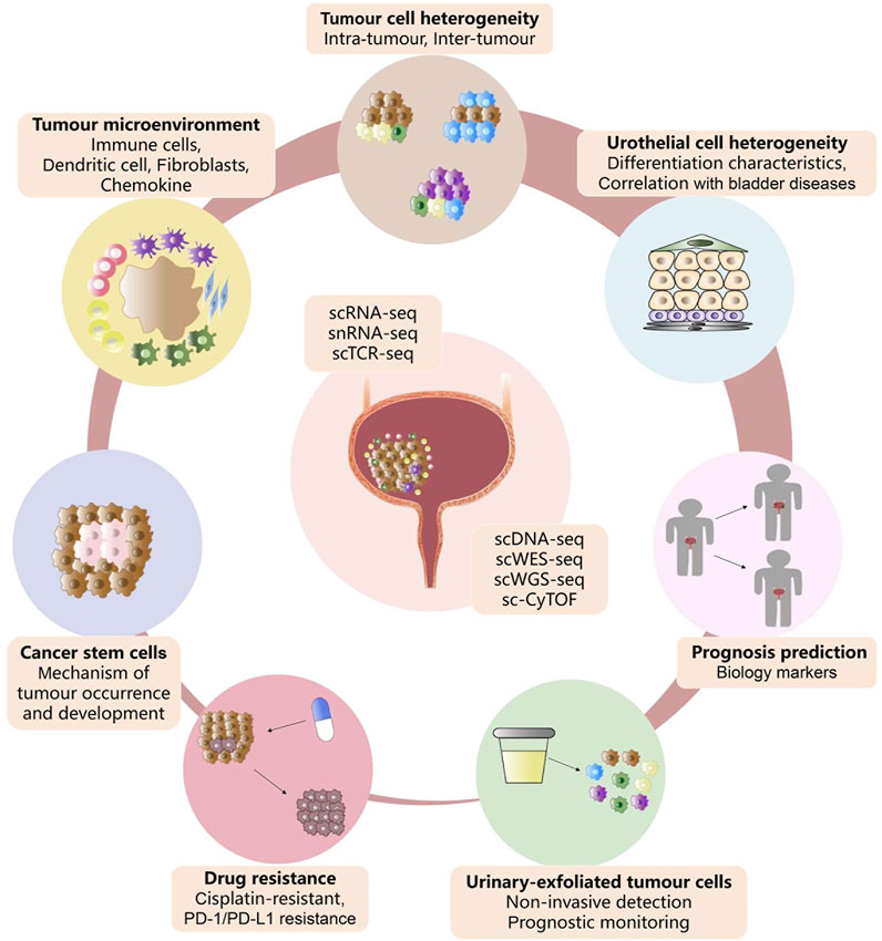
FIGURE 3. Advances of single-cell sequencing in bladder cancer. Summary of the current application of single-cell sequencing technology in the cell heterogeneity, tumor microenvironment, drug resistance, cancer stem cells, urinary-exfoliated tumor cells, and prognosis of bladder cancer.
图 3.单细胞测序在膀胱癌中的应用进展。单细胞测序技术目前在膀胱癌细胞异质性、肿瘤微环境、耐药性、癌症干细胞、尿液脱落肿瘤细胞和预后方面的应用摘要。
The past several years have witnessed unpredictable advances in single-cell sequencing technologies in the cancer field (Papalexi and Satija, 2018; Liu et al., 2021). However, the novel applications of single-cell technologies in characterising bladder cancer currently remain insufficient when compared with other human malignancies (Zang et al., 2021; Wei et al., 2022). First, existing single-cell sequencing studies have been limited to providing transcriptome or genomic information for a few specific types of bladder cancer patients, especially those lacking proteomic and cellular spatial information (Chen et al., 2018; Chen et al., 2020; Wang et al., 2020). Second, given the difficulty in obtaining the bladder urothelium of healthy individuals, single-cell sequencing studies on the link between the urothelium and bladder disease are limited to mice (Maserejian et al., 2013; Santos et al., 2019; Li et al., 2021b; Cheng et al., 2021). Third, drug resistance and metastasis often occur in bladder cancer patients, relevant single-cell research should not be limited to bladder cancer cell lines and PDX models (Tanaka et al., 2018; Lee et al., 2020). It remains indispensable to expand the study on the mechanism of bladder cancer drug resistance and metastasis through single-cell techniques in the future. As a result, our present understanding of bladder cancer cellular matrix components, TME, and drug resistance mechanisms are far from adequate. Therefore, it is a long and arduous process to comprehensively understand the underlying cellular or molecular mechanisms of bladder tumors, and plentiful studies need to be conducted.
过去几年,单细胞测序技术在癌症领域取得了不可预估的进展(Papalexi 和 Satija,2018 年;Liu 等人,2021 年)。然而,与其他人类恶性肿瘤相比,单细胞技术在表征膀胱癌方面的新应用目前仍显不足(Zang等人,2021年;Wei等人,2022年)。首先,现有的单细胞测序研究仅限于为少数特定类型的膀胱癌患者提供转录组或基因组信息,尤其是那些缺乏蛋白质组和细胞空间信息的患者(Chen等,2018;Chen等,2020;Wang等,2020)。其次,鉴于难以获得健康人的膀胱尿路上皮细胞,有关尿路上皮细胞与膀胱疾病之间联系的单细胞测序研究仅限于小鼠(Maserejian等,2013;Santos等,2019;Li等,2021b;Cheng等,2021)。第三,膀胱癌患者常出现耐药性和转移,相关的单细胞研究不应局限于膀胱癌细胞系和PDX模型(Tanaka等,2018;Lee等,2020)。未来,通过单细胞技术扩大对膀胱癌耐药和转移机制的研究仍然不可或缺。因此,我们目前对膀胱癌细胞基质成分、TME 和耐药机制的了解还远远不够。因此,全面了解膀胱肿瘤的细胞或分子机制是一个漫长而艰巨的过程,还需要开展大量的研究。
Besides, due to the complex and expensive processes, single-cell sequencing could not be applied to profile large cohorts of tumor samples (Zhang et al., 2016; Chen et al., 2018; Chen et al., 2020; Oh et al., 2020; Wang et al., 2020). Even in real-world clinics, single-cell sequencing results cannot be applied to individualized therapy due to the rapidly deteriorating health status of patients (Lee et al., 2020). Notably, limited by the algorithm, it is difficult to identify accurate proportion of rare cell subsets in batch sequencing data (Li et al., 2012; Yang et al., 2017; Chen et al., 2020). This remains a big issue in bioinformatics analysis of single-cell sequencing data, and more proofs are needed to validate these single-cell analysis results. With the ongoing technological developments, single-cell sequencing is expected to become more high-throughput, with a low cost involved, simplified data analysis process, and reduced time expenditure, that can provide better accuracy and sensitivity to collectively promote the larger-scale single-cell sequencing efforts in bladder cancer and accelerate its clinical application. Furthermore, advanced techniques such as the application of single-cell multi-omics approaches and spatial transcriptomics will help us comprehensively reveal the pathogenesis and drug resistance mechanisms of bladder cancer. With the deepening of our understanding of the cellular dynamics of bladder tumor cells, the diagnosis and treatment of bladder cancer are expected to usher in a new era of precision medicine, improve the efficacy of personalised medicine, and ultimately save the lives of bladder cancer patients.
此外,由于过程复杂且成本高昂,单细胞测序无法应用于大样本群的肿瘤图谱分析(Zhang等人,2016;Chen等人,2018;Chen等人,2020;Oh等人,2020;Wang等人,2020)。即使在现实世界的临床中,由于患者的健康状况迅速恶化,单细胞测序结果也无法应用于个体化治疗(Lee 等,2020)。值得注意的是,受限于算法,很难在批量测序数据中准确识别罕见细胞亚群的比例(李等人,2012;杨等人,2017;陈等人,2020)。这仍然是单细胞测序数据生物信息学分析中的一个大问题,需要更多的证明来验证这些单细胞分析结果。随着技术的不断发展,单细胞测序有望变得更加高通量、低成本、简化数据分析过程、减少时间支出,从而提供更好的准确性和灵敏度,共同推动膀胱癌单细胞测序工作的大规模开展,加速其临床应用。此外,单细胞多组学方法和空间转录组学等先进技术的应用将有助于我们全面揭示膀胱癌的发病机制和耐药机制。随着我们对膀胱肿瘤细胞动力学认识的加深,膀胱癌的诊断和治疗有望迎来精准医疗的新时代,提高个体化医疗的疗效,最终挽救膀胱癌患者的生命。
Author contributions 作者供稿
TL wrote the manuscript. JZ, YL, KW, and ZC took part in the manuscript modification. QZ helped with the preparation of the figure. All authors contributed to this paper and approved the submitted version.
TL 撰写了手稿。JZ、YL、KW和ZC参与了手稿的修改。QZ 协助绘制了图表。所有作者均对本文有贡献,并批准了提交的版本。
Funding 资金筹措
This work was supported by the Postdoctoral Science Foundation of China (No. 2021M690158) and the “3315 Plan” Innovation Team Project of Ningbo City (No. 2019A-14-C).
本研究得到中国博士后科学基金(编号:2021M690158)和宁波市 "3315计划 "创新团队项目(编号:2019A-14-C)的资助。
Acknowledgments 致谢
Thanks to the contributions of all authors and the support fundings in this project.
感谢所有作者的贡献以及对本项目的支持。
Conflict of interest 利益冲突
The authors declare that the research was conducted in the absence of any commercial or financial relationships that could be construed as a potential conflict of interest.
作者声明,本研究在进行过程中不存在任何可能被视为潜在利益冲突的商业或经济关系。
Publisher’s note 出版商说明
All claims expressed in this article are solely those of the authors and do not necessarily represent those of their affiliated organizations, or those of the publisher, the editors and the reviewers. Any product that may be evaluated in this article, or claim that may be made by its manufacturer, is not guaranteed or endorsed by the publisher.
本文中表述的所有主张仅代表作者本人,并不一定代表其附属机构的主张,也不代表出版商、编辑和审稿人的主张。本文可能评估的任何产品,或其制造商可能提出的任何主张,均未得到出版商的保证或认可。
References 参考资料
Antoni, S., Ferlay, J., Soerjomataram, I., Znaor, A., Jemal, A., and Bray, F. (2017). Bladder cancer incidence and mortality: A global overview and recent trends. Eur. Urol. 71, 96–108. doi:10.1016/j.eururo.2016.06.010IF: 23.4 B1
Antoni, S., Ferlay, J., Soerjomataram, I., Znaor, A., Jemal, A., and Bray, F. (2017)。膀胱癌发病率和死亡率:全球概览与最新趋势。Eur.Urol.71, 96-108. doi:10.1016/j.eururo.2016.06.010IF: 23.4 B1
PubMed Abstract | CrossRef Full Text | Google Scholar
PubMed 摘要 | CrossRef Full Text | Google Scholar
Apodaca, G. (2004). The uroepithelium: Not just a passive barrier. Traffic 5, 117–128. doi:10.1046/j.1600-0854.2003.00156.xIF: 4.5 B3
Apodaca, G. (2004).尿路上皮:不仅仅是被动屏障。doi:10.1046/j.1600-0854.2003.00156.xIF:4.5 B3
PubMed Abstract | CrossRef Full Text | Google Scholar
PubMed 摘要 | CrossRef Full Text | Google Scholar
Appels, N. M., Beijnen, J. H., and Schellens, J. H. (2005). Development of farnesyl transferase inhibitors: A review. Oncologist 10, 565–578. doi:10.1634/theoncologist.10-8-565IF: 5.8 B2
Appels, N. M., Beijnen, J. H., and Schellens, J. H. (2005).法尼基转移酶抑制剂的开发:综述。doi:10.1634/theoncologist.10-8-565IF:5.8 B2
PubMed Abstract | CrossRef Full Text | Google Scholar
PubMed 摘要 | CrossRef Full Text | Google Scholar
Audenet, F., Attalla, K., and Sfakianos, J. P. (2018). The evolution of bladder cancer genomics: What have we learned and how can we use it? Urol. Oncol. 36, 313–320. doi:10.1016/j.urolonc.2018.02.017IF: 2.7 B3
Audenet, F., Attalla, K., and Sfakianos, J. P. (2018)。膀胱癌基因组学的演变:我们学到了什么?Urol.Oncol.36, 313-320. doi:10.1016/j.urolonc.2018.02.017IF: 2.7 B3
Baras, A. S., Drake, C., Liu, J. J., Gandhi, N., Kates, M., Hoque, M. O., et al. (2016). The ratio of CD8 to Treg tumor-infiltrating lymphocytes is associated with response to cisplatin-based neoadjuvant chemotherapy in patients with muscle invasive urothelial carcinoma of the bladder. OncoImmunology 5, e1134412. doi:10.1080/2162402X.2015.1134412IF: 7.2 B2
Batista, R., Vinagre, N., Meireles, S., Vinagre, J., Prazeres, H., Leão, R., et al. (2020). Biomarkers for bladder cancer diagnosis and surveillance: A comprehensive review. Diagn. (Basel) 10, 39. doi:10.3390/diagnostics10010039IF: 3.6 B3
Berdik, C. (2017). Unlocking bladder cancer. Nature 551, S34–S35. S35. doi:10.1038/551S34aIF: 64.8 B1
Bryan, R. T. (2015). Cell adhesion and urothelial bladder cancer: The role of cadherin switching and related phenomena. Philos. Trans. R. Soc. Lond. B Biol. Sci. 370, 20140042. doi:10.1098/rstb.2014.0042IF: 6.3 B2
Burger, M., Catto, J. W., Dalbagni, G., Grossman, H. B., Herr, H., Karakiewicz, P., et al. (2013). Epidemiology and risk factors of urothelial bladder cancer. Eur. Urol. 63, 234–241. doi:10.1016/j.eururo.2012.07.033IF: 23.4 B1
Cao, R., Yuan, L. S., Ma, B., Wang, G., Qiu, W., and Tian, Y. (2020). An EMT-related gene signature for the prognosis of human bladder cancer. J. Cell. Mol. Med. 24, 605–617. doi:10.1111/jcmm.14767IF: 5.3 B2
Casadei, C., Dizman, N., Schepisi, G., Cursano, M. C., Basso, U., Santini, D., et al. (2019). Targeted therapies for advanced bladder cancer: New strategies with FGFR inhibitors. Ther. Adv. Med. Oncol. 11, 1758835919890285. doi:10.1177/1758835919890285IF: 4.9 B2
Castillo-Martin, M., Domingo-Domenech, J., Karni-Schmidt, O., Matos, T., and Cordon-Cardo, C. (2010). Molecular pathways of urothelial development and bladder tumorigenesis. Urol. Oncol. 28, 401–408. doi:10.1016/j.urolonc.2009.04.019IF: 2.7 B3
Chen, A., Fu, G., Xu, Z., Sun, Y., Chen, X., Cheng, K., et al. (2018). Detection of urothelial bladder carcinoma via microfluidic immunoassay and single-cell DNA copy-number alteration analysis of captured urinary-exfoliated tumor cells. Cancer Res. 78, 4073–4085. doi:10.1158/0008-5472.CAN-17-2615IF: 11.2 B1
Chen, Y. P., Zhang, Y., Lv, J. W., Li, Y. Q., Wang, Y. Q., He, Q. M., et al. (2017). Genomic analysis of tumor microenvironment immune types across 14 solid cancer types: Immunotherapeutic implications. Theranostics 7, 3585–3594. doi:10.7150/thno.21471IF: 12.4 B1
Chen, Z., Zhou, L., Liu, L., Hou, Y., Xiong, M., Yang, Y., et al. (2020). Single-cell RNA sequencing highlights the role of inflammatory cancer-associated fibroblasts in bladder urothelial carcinoma. Nat. Commun. 11 (1), 5077. doi:10.1038/s41467-020-18916-5IF: 16.6 B1
Cheng, X., Lai, H., Luo, W., Zhang, M., Miao, J., Song, W., et al. (2021). Single-cell analysis reveals urothelial cell heterogeneity and regenerative cues following cyclophosphamide-induced bladder injury. Cell Death Dis. 12, 446. doi:10.1038/s41419-021-03740-6IF: 9.0 B1
Compérat, E. M., Burger, M., Gontero, P., Mostafid, A. H., Palou, J., Rouprêt, M., et al. (2019). Grading of urothelial carcinoma and the new “World Health Organisation classification of tumours of the urinary system and male genital organs 2016”. Eur. Urol. Focus 5, 457–466. doi:10.1016/j.euf.2018.01.003IF: 5.4 B2
Critelli, R., Fasanelli, F., Oderda, M., Polidoro, S., Assumma, M. B., Viberti, C., et al. (2016). Detection of multiple mutations in urinary exfoliated cells from male bladder cancer patients at diagnosis and during follow-up. Oncotarget 7, 67435–67448. doi:10.18632/oncotarget.11883
Feng, C., Wang, X., Tao, Y., Xie, Y., Lai, Z., Li, Z., et al. (2021). Single-cell proteomic analysis dissects the complexity of tumor microenvironment in muscle invasive bladder cancer. Cancers (Basel) 13 (21), 5440. doi:10.3390/cancers13215440IF: 5.2 B2
Gouin, K. H., Ing, N., Plummer, J. T., Rosser, C., Cheikh, B., Oh, C., et al. (2021). An N-Cadherin 2 expressing epithelial cell subpopulation predicts response to surgery, chemotherapy and immunotherapy in bladder cancer. Nat. Commun. 12 (1), 4906. doi:10.1038/s41467-021-25103-7IF: 16.6 B1
Guo, X., Zhang, Y., Zheng, L., Zheng, C., Song, J., Zhang, Q., et al. (2018). Global characterization of T cells in non-small-cell lung cancer by single-cell sequencing. Nat. Med. 24, 978–985. doi:10.1038/s41591-018-0045-3IF: 82.9 B1
Hargadon, K. M., Johnson, C. E., and Williams, C. J. (2018). Immune checkpoint blockade therapy for cancer: An overview of FDA-approved immune checkpoint inhibitors. Int. Immunopharmacol. 62, 29–39. doi:10.1016/j.intimp.2018.06.001IF: 5.6 B2
Hatogai, K., and Sweis, R. F. (2020). The tumor microenvironment of bladder cancer. Adv. Exp. Med. Biol. 1296, 275–290. doi:10.1007/978-3-030-59038-3_17
Heide, T., Maurer, A., Eipel, M., Knoll, K., Geelvink, M., Veeck, J., et al. (2019). Multiregion human bladder cancer sequencing reveals tumour evolution, bladder cancer phenotypes and implications for targeted therapy. J. Pathol. 248, 230–242. doi:10.1002/path.5250IF: 7.3 B2
Heng, H. H., Stevens, J. B., Liu, G., Bremer, S. W., Ye, K. J., Reddy, P. V., et al. (2006). Stochastic cancer progression driven by non-clonal chromosome aberrations. J. Cell. Physiol. 208 (2), 461–472. doi:10.1002/jcp.20685IF: 5.6 B2
Ho, P. L., Kurtova, A., and Chan, K. S. (2012). Normal and neoplastic urothelial stem cells: Getting to the root of the problem. Nat. Rev. Urol. 9, 583–594. doi:10.1038/nrurol.2012.142IF: 15.3 B1
Huang, R., Wang, L., He, J., and Gao, W. (2021). Application and prospects of single cell sequencing in tumors. Biomark. Res. 9 (1), 88. doi:10.1186/s40364-021-00336-2IF: 11.1 B2
Junttila, M. R., and de Sauvage, F. J. (2013). Influence of tumour micro-environment heterogeneity on therapeutic response. Nature 501, 346–354. doi:10.1038/nature12626IF: 64.8 B1
Kamoun, A., de Reyniès, A., Allory, Y., Sjödahl, G., Robertson, A. G., Seiler, R., et al. (2019). A consensus molecular classification of muscle-invasive bladder cancer. Eur. Urol. 77, 420–433. doi:10.1016/j.eururo.2019.09.006IF: 23.4 B1
Kantor, A. F., Hartge, P., Hoover, R. N., and Fraumeni, J. F. (1988). Epidemiological characteristics of squamous cell carcinoma and adenocarcinoma of the bladder. Cancer Res. 48, 3853–3855.
Kashima, Y., Sakamoto, Y., Kaneko, K., Seki, M., Suzuki, Y., and Suzuki, A. (2020). Single-cell sequencing techniques from individual to multiomics analyses. Exp. Mol. Med. 52 (9), 1419–1427. doi:10.1038/s12276-020-00499-2IF: 12.8 B2
Kerzeli, I. K., Lord, M., Doroszko, M., Elgendy, R., Chourlia, A., Stepanek, I., et al. (2021). Single-cell RNAseq and longitudinal proteomic analysis of a novel semi-spontaneous urothelial cancer model reveals tumor cell heterogeneity and pretumoral urine protein alterations. PLoS One 16, 0253178. doi:10.1371/journal.pone.0253178IF: 3.7 B3
Khandelwal, P., Abraham, S. N., and Apodaca, G. (2009). Cell biology and physiology of the uroepithelium. Am. J. Physiol. Ren. Physiol. 297, 1477–1501. doi:10.1152/ajprenal.00327.2009IF: 4.2 B2
Knowles, M., and Hurst, C. (2015). Molecular biology of bladder cancer: New insights into pathogenesis and clinical diversity. Nat. Rev. Cancer 15, 25–41. doi:10.1038/nrc3817IF: 78.5 B1
Lai, H., Cheng, X., Liu, Q., Luo, W., Liu, M., Zhang, M., et al. (2021). Single-cell RNA sequencing reveals the epithelial cell heterogeneity and invasive subpopulation in human bladder cancer. Int. J. Cancer 149, 2099–2115. doi:10.1002/ijc.33794IF: 6.4 B2
Lee, H. W., Chung, W., Lee, H. O., Jeong, D. E., Jo, A., Lim, J. E., et al. (2020). Single-cell RNA sequencing reveals the tumor microenvironment and facilitates strategic choices to circumvent treatment failure in a chemorefractory bladder cancer patient. Genome Med. 12, 47. doi:10.1186/s13073-020-00741-6IF: 12.3 B1
Lei, Y., Tang, R., Xu, J., Wang, W., Zhang, B., Liu, J., et al. (2021). Applications of single-cell sequencing in cancer research: Progress and perspectives. J. Hematol. Oncol. 14, 91. doi:10.1186/s13045-021-01105-2IF: 28.5 B1
Lerner, S. P., McConkey, D. J., Hoadley, K. A., Chan, K. S., Kim, W. Y., Radvanyi, F., et al. (2016). Bladder cancer molecular taxonomy: Summary from a consensus meeting. Bladder Cancer 2, 37–47. doi:10.3233/BLC-150037IF: 1.1 B4
Letašiová, S., Medve'ová, A., Šovčíková, A., Dušinská, M., Volkovová, K., Mosoiu, C., et al. (2012). Bladder cancer, a review of the environmental risk factors. Environ. Health 11, 11. doi:10.1186/1476-069X-11-S1-S11IF: 6.0 B2
Li, D. Y., Yang, F., Liao, W. Q., Zhou, X. F., Li, W. B., Cai, J. R., et al. (2021). Deep genomic sequencing of bladder urothelial carcinoma in southern Chinese patients: A single-center study. Front. Oncol. 11, 538927. doi:10.3389/fonc.2021.538927IF: 4.7 B3
Li, Y., Liu, Y., Gao, Z., Zhang, L., Chen, L., Wu, Z., et al. (2021). Single-cell transcriptomes of mouse bladder urothelium uncover novel cell type markers and urothelial differentiation characteristics. Cell Prolif. 54, 13007. doi:10.1111/cpr.13007IF: 8.5 B1
Li, Y., Xu, X., Song, L., Hou, Y., Li, Z., Tsang, S., et al. (2012). Single-cell sequencing analysis characterizes common and cell-lineage-specific mutations in a muscle-invasive bladder cancer. Gigascience 1, 12. doi:10.1186/2047-217X-1-12IF: 9.2 B2
Ligon, M. M., Wang, C., DeJong, E. N., Schulz, C., Bowdish, D., and Mysorekar, I. (2020). Single cell and tissue-transcriptomic analysis of murine bladders reveals age- and TNFα-dependent but microbiota-independent tertiary lymphoid tissue formation. Mucosal Immunol. 13, 908–918. doi:10.1038/s41385-020-0290-xIF: 8.0 B2
Lin, J., Spitz, M. R., Dinney, C. P., Etzel, C. J., Grossman, H. B., and Wu, X. (2006). Bladder cancer risk as modified by family history and smoking. Cancer 107, 705–711. doi:10.1002/cncr.22071IF: 6.2 B2
Lin, X., Deng, T., Wu, S., Lin, S., Wang, D., Wu, C-L., et al. (2019). The clinicopathological characteristics and prognostic value of squamous differentiation in patients with bladder urothelial carcinoma: A meta-analysis. World J. Urol. 38 (2), 323–333. doi:10.1007/s00345-019-02771-1IF: 3.4 B2
Liu, J., Qu, S., Zhang, T., Gao, Y., Shi, H., Song, K., et al. (2021). Applications of single-cell omics in tumor immunology. Front. Immunol. 12, 697412. doi:10.3389/fimmu.2021.697412IF: 7.3 B2
Maserejian, N. N., Chen, S., Chiu, G. R., Wager, C. G., Kupelian, V., Araujo, A. B., et al. (2013). Incidence of lower urinary tract symptoms in a population-based study of men and women. Urology 82 (3), 560–564. doi:10.1016/j.urology.2013.05.009IF: 2.1 B3
Minato, A., Noguchi, H., Tomisaki, I., Fukuda, A., Kubo, T., Nakayama, T., et al. (2018). Clinical significance of squamous differentiation in urothelial carcinoma of the bladder. Cancer control. 25, 1073274818800269. doi:10.1177/1073274818800269IF: 2.6 B4
Minoli, M., Kiener, M., Thalmann, G. N., Kruithof-de Julio, M., and Seiler, R. (2020). Evolution of urothelial bladder cancer in the context of molecular classifications. Int. J. Mol. Sci. 21, 5670. doi:10.3390/ijms21165670IF: 5.6 B2
Oh, D. Y., Kwek, S. S., Raju, S. S., Li, T., McCarthy, A., Chow, E., et al. (2020). Intratumoral CD4+ T cells mediate anti-tumor cytotoxicity in human bladder cancer. Cell 181, 1612–1625. e13. doi:10.1016/j.cell.2020.05.017IF: 64.5 B1
Papafotiou, G., Paraskevopoulou, V., Vasilaki, E., Kanaki, Z., Paschalidis, N., and Klinakis, A. (2016). KRT14 marks a subpopulation of bladder basal cells with pivotal role in regeneration and tumorigenesis. Nat. Commun. 7, 11914. doi:10.1038/ncomms11914IF: 16.6 B1
Papalexi, E., and Satija, R. (2018). Single-cell RNA sequencing to explore immune cell heterogeneity. Nat. Rev. Immunol. 18, 35–45. doi:10.1038/nri.2017.76IF: 100.3 B1
Pietzak, E. J., Bagrodia, A., Cha, E. K., Drill, E. N., Iyer, G., Isharwal, S., et al. (2017). Next-generation sequencing of nonmuscle invasive bladder cancer reveals potential biomarkers and rational therapeutic targets. Eur. Urol. 72, 952–959. doi:10.1016/j.eururo.2017.05.032IF: 23.4 B1
Pitzalis, C., Jones, G. W., Bombardieri, M., and Jones, S. A. (2014). Ectopic lymphoid-like structures in infection, cancer and autoimmunity. Nat. Rev. Immunol. 14, 447–462. doi:10.1038/nri3700IF: 100.3 B1
Ren, X., Kang, B., and Zhang, Z. (2018). Understanding tumor ecosystems by single-cell sequencing: Promises and limitations. Genome Biol. 19, 211. doi:10.1186/s13059-018-1593-zIF: 12.3 B1
Roy, S., Pradhan, D., Ernst, W. L., Mercurio, S., Najjar, Y., Parikh, R., et al. (2017). Next-generation sequencing-based molecular characterization of primary urinary bladder adenocarcinoma. Mod. Pathol. 30, 1133–1143. doi:10.1038/modpathol.2017.33IF: 7.5 B1
Santos, C. P., Lapi, E., Martínez de Villarreal, J., Álvaro-Espinosa, L., Fernández-Barral, A., Barbáchano, A., et al. (2019). Urothelial organoids originating from Cd49fhigh mouse stem cells display Notch-dependent differentiation capacity. Nat. Commun. 10, 4407. doi:10.1038/s41467-019-12307-1IF: 16.6 B1
Saunders, N. A., Simpson, F., Thompson, E. W., Hill, M. M., Endo-Munoz, L., Leggatt, G., et al. (2012). Role of intratumoural heterogeneity in cancer drug resistance: Molecular and clinical perspectives. EMBO Mol. Med. 4, 675–684. doi:10.1002/emmm.201101131IF: 11.1 B1
Sfakianos, J. P., Daza, J., Yang, H., Anastos, H., Bryant, G., Bareja, R., et al. (2020). Epithelial plasticity can generate multi-lineage phenotypes in human and murine bladder cancers. Nat. Commun. 11, 2540. doi:10.1038/s41467-020-16162-3IF: 16.6 B1
Shin, K., Lee, J., Guo, N., Kim, J., Lim, A., Qu, L., et al. (2011). Hedgehog/Wnt feedback supports regenerative proliferation of epithelial stem cells in bladder. Nature 472, 110–114. doi:10.1038/nature09851IF: 64.8 B1
Sjödahl, G., Eriksson, P., Liedberg, F., and Höglund, M. (2017). Molecular classification of urothelial carcinoma: Global mRNA classification versus tumour-cell phenotype classification. J. Pathol. 242, 113–125. doi:10.1002/path.4886IF: 7.3 B2
Sjödahl, G., Jackson, C. L., Bartlett, J. M., Siemens, D. R., and Berman, D. M. (2019). Molecular profiling in muscle-invasive bladder cancer: More than the sum of its parts. J. Pathol. 247, 563–573. doi:10.1002/path.5230IF: 7.3 B2
Song, D., Powles, T., Shi, L., Zhang, L., Ingersoll, , and Lu, Y. (2019). Bladder cancer, a unique model to understand cancer immunity and develop immunotherapy approaches. J. Pathol. 249 (2), 151–165. doi:10.1002/path.5306IF: 7.3 B2
Stuart, T., and Satija, R. (2019). Integrative single-cell analysis. Nat. Rev. Genet. 20, 257–272. doi:10.1038/s41576-019-0093-7IF: 42.7 B1
Tanaka, N., Katayama, S., Reddy, A., Nishimura, K., Niwa, N., Hongo, H., et al. (2018). Single‐cell RNA‐seq analysis reveals the platinum resistance gene COX7B and the surrogate marker CD63. Cancer Med. 7, 6193–6204. doi:10.1002/cam4.1828IF: 4.0 B2
Tang, F., Barbacioru, C., Wang, Y., Nordman, E., Lee, C., Xu, N., et al. (2009). mRNA-seq whole-transcriptome analysis of a single cell. Nat. Methods 6, 377–382. doi:10.1038/nmeth.1315IF: 48.0 B1
Thomsen, M. B. H., Nordentoft, I., Lamy, P., Vang, S., Reinert, L., Mapendano, C. K., et al. (2017). Comprehensive multiregional analysis of molecular heterogeneity in bladder cancer. Sci. Rep. 7, 11702. doi:10.1038/s41598-017-11291-0IF: 4.6 B2
Tran, L., Xiao, J. F., Agarwal, N., Duex, J. E., and Theodorescu, D. (2021). Advances in bladder cancer biology and therapy. Nat. Rev. Cancer 21, 104–121. doi:10.1038/s41568-020-00313-1IF: 78.5 B1
van den Bosch, S., and Alfred Witjes, J. (2011). Long-term cancer-specific survival in patients with high-risk, non-muscle-invasive bladder cancer and tumour progression: A systematic review. Eur. Urol. 60, 493–500. doi:10.1016/j.eururo.2011.05.045IF: 23.4 B1
Wang, H., Mei, Y., Luo, C., Huang, Q., Wang, Z., Lu, G., et al. (2021). Single-cell analyses reveal mechanisms of cancer stem cell maintenance and epithelial-mesenchymal transition in recurrent bladder cancer. Clin. Cancer Res. 27 (22), 6265–6278. doi:10.1158/1078-0432.CCR-20-4796IF: 11.5 B1
Wang, J., Batourina, E., Schneider, K., Souza, S., Swayne, T., Liu, C., et al. (2018). Polyploid superficial cells that maintain the urothelial barrier are produced via incomplete cytokinesis and endoreplication. Cell Rep. 25, 464–477. e4. doi:10.1016/j.celrep.2018.09.042IF: 8.8 B1
Wang, J., Yao, X., and Huang, J. (2017). New tricks for human farnesyltransferase inhibitor: Cancer and beyond. Medchemcomm 8, 841–854. doi:10.1039/c7md00030h
Wang, L., Sfakianos, J. P., Beaumont, K. G., Akturk, G., Horowitz, A., Sebra, R. P., et al. (2021). Myeloid cell-associated resistance to PD-1/PD-L1 blockade in urothelial cancer revealed through bulk and single-cell RNA sequencing. Clin. Cancer Res. 27 (15), 4287–4300. doi:10.1158/1078-0432.CCR-20-4574IF: 11.5 B1
Wang, P. F., Nishitani, M. A., Tanimoto, S., Kishimoto, T., Fukumori, T., Takahashi, M., et al. (2007). Bladder cancer cell invasion is enhanced by cross-talk with fibroblasts through hepatocyte growth factor. Urology 69, 780–784. doi:10.1016/j.urology.2007.01.063IF: 2.1 B3
Wang, X., Pan, L., Lu, Q., Huang, H., Feng, C., Tao, Y., et al. (2021). A combination of ssGSEA and mass cytometry identifies immune microenvironment in muscle-invasive bladder cancer. J. Clin. Lab. Anal. 35 (5), e23754. doi:10.1002/jcla.23754IF: 2.7 B4
Wang, Z., Chen, J., Yang, L., Cao, M., Yu, Y., Zhang, R., et al. (2020). Single-cell sequencing-enabled Hexokinase 2 assay for noninvasive bladder cancer diagnosis and screening by detecting rare malignant cells in urine. Anal. Chem. 92, 16284–16292. doi:10.1021/acs.analchem.0c04282IF: 7.4 B1
Wei, W., Rong, Y., Sanhe, L., Chunxiu, Y., Thokerunga, E., Cui, D., et al. (2022). Single-cell sequencing and its applications in bladder cancer. Expert Rev. Mol. Med. 24, e6. doi:10.1017/erm.2021.23IF: 6.2 B2
Witjes, J. A., Bruins, H. M., Cathomas, R., Compérat, E. M., Cowan, N. C., Gakis, G., et al. (2020). European association of urology guidelines on muscle-invasive and metastatic bladder cancer: Summary of the 2020 guidelines. Eur. Urol. 79, 82–104. doi:10.1016/j.eururo.2020.03.055IF: 23.4 B1
Wu, S., Ou, T., Xing, N., Lu, J., Wan, S., Wang, C., et al. (2019). Whole-genome sequencing identifies ADGRG6 enhancer mutations and FRS2 duplications as angiogenesis-related drivers in bladder cancer. Nat. Commun. 10, 720. doi:10.1038/s41467-019-08576-5IF: 16.6 B1
Wu, X., Lv, D., Cai, C., Zhao, Z., Wang, M., Chen, W., et al. (2020). A TP53-associated immune prognostic signature for the prediction of overall survival and therapeutic responses in muscle-invasive bladder cancer. Front. Immunol. 11, 590618. doi:10.3389/fimmu.2020.590618IF: 7.3 B2
Xing, Q. R., Cipta, N. O., Hamashima, K., Liou, Y. C., Koh, C. G., and Loh, Y. H. (2020). Unraveling heterogeneity in transcriptome and its regulation through single-cell multi-omics technologies. Front. Genet. 11, 662. doi:10.3389/fgene.2020.00662IF: 3.7 B3
Xu, S., Catapang, A., Braas, D., Stiles, L., Doh, H., Lee, J., et al. (2018). A precision therapeutic strategy for hexokinase 1-null, hexokinase 2-positive cancers. Cancer Metab. 6, 7. doi:10.1186/s40170-018-0181-8IF: 5.9 B3
Yafi, F. A., Brimo, F., Steinberg, J., Aprikian, A. G., Tanguay, S., and Kassouf, W. (2015). Prospective analysis of sensitivity and specificity of urinary cytology and other urinary biomarkers for bladder cancer. Urol. Oncol. 33, 6625–e31. doi:10.1016/j.urolonc.2014.06.008IF: 2.7 B3
Yang, Z., Li, C., Fan, Z., Liu, H., Zhang, X., Cai, Z., et al. (2017). Single-cell sequencing reveals variants in ARID1A, GPRC5A and MLL2 driving self-renewal of human bladder cancer stem cells. Eur. Urol. 71 (1), 8–12. doi:10.1016/j.eururo.2016.06.025IF: 23.4 B1
Yu, Z., Liao, J., Chen, Y., Zhang, L., Zou, C., Zhang, H., et al. (2019). Single-cell transcriptomic map of the human and mouse bladders. J. Am. Soc. Nephrol. 30, 2159–2176. doi:10.1681/ASN.2019040335IF: 13.6 B1
Zang, J., Ye, K., Fei, Y., Zhang, R., Chen, H., and Zhuang, G. (2021). Immunotherapy in the treatment of urothelial bladder cancer: Insights from single-cell analysis. Front. Oncol. 11, 696716. doi:10.3389/fonc.2021.696716IF: 4.7 B3
Zhang, X., Meng, Z., Yong, H., Xu, L., Li, W., Zou, Z., et al. (2016). Single-cell analyses of transcriptional heterogeneity in squamous cell carcinoma of urinary bladder. Oncotarget 7 (40), 66069–66076. doi:10.18632/oncotarget.11803
Zhang, Y., and Zhang, Z. (2020). The history and advances in cancer immunotherapy: Understanding the characteristics of tumor-infiltrating immune cells and their therapeutic implications. Cell. Mol. Immunol. 17, 807–821. doi:10.1038/s41423-020-0488-6IF: 24.1 B1
Zheng, C., Zheng, L., Yoo, J. K., Guo, H., Zhang, Y., Guo, X., et al. (2017). Landscape of infiltrating T cells in liver cancer revealed by single-cell sequencing. Cell 169, 1342–1356. e16. doi:10.1016/j.cell.2017.05.035IF: 64.5 B1
Zhou, R., Liang, J., Chen, Q., Tian, H., Yang, C., and Liu, C. (2021). Development and validation of an intra-tumor heterogeneity-related signature to predict prognosis of bladder cancer: A study based on single-cell RNA-seq. Aging 13, 19415–19441. doi:10.18632/aging.203353IF: 5.2 B3
Zhu, S., Yu, W., Yang, X., Wu, C., and Cheng, F. (2020). Traditional classification and novel subtyping systems for bladder cancer. Front. Oncol. 10, 102. doi:10.3389/fonc.2020.00102IF: 4.7 B3
Keywords: single-cell sequencing, bladder cancer, heterogeneity, biomarkers, precision medicine
Citation: Lyu T, Lin Y, Wu K, Cao Z, Zhang Q and Zheng J (2022) Single-cell sequencing technologies in bladder cancer research: Applications and challenges. Front. Genet. 13:1027909. doi: 10.3389/fgene.2022.1027909IF: 3.7 B3
Received: 25 August 2022; Accepted: 06 October 2022;
Published: 19 October 2022.
Edited by:
Sathiya Pandi Narayanan, Sidra Medicine, QatarReviewed by:
Liangkuan Bi, Second Affiliated Hospital of Anhui Medical University, ChinaHung N. Luu, University of Pittsburgh, United States
Copyright © 2022 Lyu, Lin, Wu, Cao, Zhang and Zheng. This is an open-access article distributed under the terms of the Creative Commons Attribution License (CC BY). The use, distribution or reproduction in other forums is permitted, provided the original author(s) and the copyright owner(s) are credited and that the original publication in this journal is cited, in accordance with accepted academic practice. No use, distribution or reproduction is permitted which does not comply with these terms.
*Correspondence: Jianping Zheng, zhengjianping@nimte.ac.cn