Abstract 抽象
The protein disulfide isomerase (PDI) family is a group of thioredoxin endoplasmic reticulum (ER)-resident enzymes and molecular chaperones that play crucial roles in the correct folding of proteins. PDIs are upregulated in multiple cancer types and are considered a novel target for cancer therapy. In this study, we found that a potent pan-PDI inhibitor, E64FC26, significantly decreased the proliferation of pancreatic ductal adenocarcinoma (PDAC) cells. As expected, E64FC26 treatment increased ER stress and the unfolded protein response (UPR), as evidenced by upregulation of glucose-regulated protein, 78-kDa (GRP78), phosphorylated (p)-PKR-like ER kinase (PERK), and p-eukaryotic initiation factor 2α (eIF2α). Persistent ER stress was found to lead to apoptosis, ferroptosis, and autophagy, all of which are dependent on lysosomal functions. First, there was little cleaved caspase-3 in E64FC26-treated cells according to Western blotting, but a higher dose of E64FC26 was needed to induce caspase activity. Then, E64FC26-induced cell death could be reversed by adding the iron chelator, deferoxamine, and the reactive oxygen species scavengers, ferrostatin-1 and N-acetylcysteine. Furthermore, the autophagosome-specific marker, light chain 3B (LC3B)-II, increased, but the autolysosome marker, sequestosome 1 (SQSTM1)/p62, was not degraded in E64FC26-treated cells. Using the FUW mCherry-LC3 plasmid and acridine orange staining, we also discovered a lower number of acidic vesicles, such as autolysosomes and mature lysosomes, in E64FC26-treated cells. Finally, E64FC26 treatment increased the cathepsin L precursor (pre-CTSL) but decreased mature CTSL expression according to Western blotting, indicating a defective lysosome. These results suggested that the PDI inhibitor, E64FC26, might initially impede proper folding of proteins, and then induce ER stress and disrupt proteostasis, subsequently leading to lysosomal defects. Due to defective lysosomes, the extents of apoptosis and ferroptosis were limited, and fusion with autophagosomes was blocked in E64FC26-treated cells. Blockade of autolysosomal formation further led to the autophagic cell death of PDAC cells.
蛋白质二硫键异构酶 (PDI) 家族是一组硫氧还蛋白内质网 (ER) 驻留酶和分子伴侣,在蛋白质的正确折叠中起着至关重要的作用。PDI 在多种癌症类型中上调,被认为是癌症治疗的新靶点。在这项研究中,我们发现一种有效的泛 PDI 抑制剂 E64FC26 显着降低了胰导管腺癌 (PDAC) 细胞的增殖。正如预期的那样,E64FC26处理增加了 ER 应激和未折叠蛋白反应 (UPR),葡萄糖调节蛋白 78-kDa (GRP78) 、磷酸化 (p)-PKR 样 ER 激酶 (PERK) 和 p-真核起始因子 2α (eIF2α) 的上调证明了这一点。发现持续的 ER 应激导致细胞凋亡、铁死亡和自噬,所有这些都依赖于溶酶体功能。首先,根据 Western blotting,E64FC26处理的细胞中几乎没有裂解的 caspase-3,但需要更高剂量的 E64FC26 来诱导 caspase 活性。然后,通过添加铁螯合剂、去铁胺和活性氧清除剂、ferrostatin-1 和 N-乙酰半胱氨酸,可以逆转 E64FC26 诱导的细胞死亡。此外,自噬体特异性标志物轻链 3B (LC3B)-II 增加,但自噬溶酶体标志物 sequestosome 1 (SQSTM1)/p62 在 E64FC26 处理的细胞中没有降解。使用 FUW mCherry-LC3 质粒和吖啶橙染色,我们还发现 E64FC26 处理的细胞中酸性囊泡(如自溶酶体和成熟溶酶体)数量较少。最后,根据 Western blotting,E64FC26处理增加了组织蛋白酶 L 前体 (pre-CTSL),但降低了成熟 CTSL 的表达,表明溶酶体有缺陷。 这些结果表明,PDI 抑制剂 E64FC26 最初可能阻碍蛋白质的正确折叠,然后诱导 ER 应激并破坏蛋白质稳态,随后导致溶酶体缺陷。由于溶酶体缺陷,细胞凋亡和铁死亡的范围受到限制,并且在 E64FC26 处理的细胞中与自噬体的融合被阻断。阻断自噬溶酶体形成进一步导致 PDAC 细胞自噬细胞死亡。
Keywords: protein disulfide isomerase, ferroptosis, apoptosis, autophagy, pancreatic ductal adenocarcinoma
关键字:蛋白二硫键异构酶, 铁死亡, 细胞凋亡, 自噬, 胰腺导管腺癌
1. Introduction 1. 引言
In eukaryotic cells, the endoplasmic reticulum (ER) is responsible for protein folding and post-translational modifications, and it stores calcium ions [1]. As is known, mitochondria are the main organelles that produce reactive oxygen species (ROS) in cells. However, large amounts of ROS are also produced during oxidative protein folding to form disulfide bonds [2,3]. The main enzymes responsible for the formation of disulfide bonds in nascent proteins in the ER are members of the protein disulfide isomerase (PDI) family and the ER oxidoreductase 1 (ERO1) family. They are all thiol oxidoreductases that mainly remain in the ER [2,3]. The active cysteine site in PDIs usually accepts electrons from the free thiol of nascent proteins, so nascent proteins can form disulfide bonds. Afterward, PDI transfers electrons to ERO1, and ERO1 further transfers electrons to O2, thereby generating H2O2. Under normal situations, the H2O2 can be converted into water and oxygen by many antioxidant enzymes, including superoxide dismutase, catalase, ascorbate peroxidase, glutathione peroxidases, and peroxiredoxin 4, or eliminated by ROS scavengers, such as glutathione and vitamin E [4,5].
在真核细胞中,内质网 (ER) 负责蛋白质折叠和翻译后修饰,并储存钙离子 [ 1 ]。众所周知,线粒体是在细胞中产生活性氧 (ROS) 的主要细胞器。然而,在氧化蛋白质折叠形成二硫键的过程中也会产生大量的 ROS [ 2 , 3 ]。负责在 ER 中新生蛋白质中形成二硫键的主要酶是蛋白质二硫键异构酶 (PDI) 家族和 ER 氧化还原酶 1 (ERO1) 家族的成员。它们都是主要保留在 ER 中的 巯基氧化还原酶 [ 2 , 3 ]。PDI 中的活性半胱氨酸位点通常接受来自新生蛋白质的游离硫醇的电子,因此新生蛋白质可以形成二硫键。之后,PDI 将电子转移到 ERO1,ERO1 进一步将电子转移到 O2,从而产生 H2O2。在正常情况下,H2O2 可被许多抗氧化酶转化为水和氧气,包括超氧化物歧化酶、过氧化氢酶、抗坏血酸过氧化物酶、谷胱甘肽过氧化物酶和过氧化物还原蛋白 4,或被 ROS 清除剂(如谷胱甘肽和维生素 E)消除 [ 4 , 5 ]。
PDI family members are ubiquitously expressed in various tissues and cell types in mammals, are especially abundant in secretory tissues, and play vital roles in protein folding and maintaining normal cell functions. Misfolding of proteins can affect their normal biological functions, and the accumulation of misfolded proteins also causes related diseases [6], such as neurodegenerative disorders [7], pulmonary emphysema [8], and cystic fibrosis [9]. In recent years, it was found that PDI family members are overexpressed in various cancers [10], and PDI family members contribute to tumor proliferation and malignancy, and are related to resistance to chemotherapeutic drugs. In most cases, high expression levels of PDI family members have a protective effect on cancer cells and are associated with poor patient survival [11]. In breast cancer stem cells, knockdown of PDI family members, such as PDIA1, ER resident protein 44 (ERp44), or ERp57, inhibits cell proliferation [12], and overexpressions of PDI family members, such as PDIA3 and PDIA6, are associated with invasiveness of primary ductal breast cancer [13]. High expression of anterior gradient protein 2 homolog (AGR2), a member of the PDI family, is related to low survival of lung cancer patients [14]. Among PDI family members, PDIA1 is the most critical molecule involved in protein folding and is associated with a variety of diseases, including cancers [15], thrombosis [16], Alzheimer’s disease [17], and diabetes [18]. PDIA1 is overexpressed in a variety of cancers to meet the growing proliferation needs of tumors. Inhibition of PDIA1 by PACMA31 causes toxicity in ovarian cancer cells [19]. Therefore, the development of PDI inhibitors for cancer treatment has become an important topic in recent years [20]. Currently, many PDI inhibitors have been developed; however, those drugs are still in preclinical or early clinical research stages.
PDI 家族成员在哺乳动物的各种组织和细胞类型中普遍表达,在分泌组织中尤其丰富,在蛋白质折叠和维持正常细胞功能中起着至关重要的作用。蛋白质的错误折叠会影响其正常的生物学功能,错误折叠的蛋白质的积累也会引起相关疾病 [ 6 ],如神经退行性疾病 [ 7 ]、肺气肿 [ 8 ] 和囊性纤维化 [ 9 ]。近年来,发现 PDI 家族成员在各种癌症中过表达 [ 10 ],PDI 家族成员导致肿瘤增殖和恶性肿瘤,并与化疗药物耐药有关。在大多数情况下,PDI 家族成员的高表达水平对癌细胞具有保护作用,并且与患者生存率差有关 [ 11 ]。在乳腺癌干细胞中,敲除 PDI 家族成员,如 PDIA1、ER 驻留蛋白 44 (ERp44) 或 ERp57,抑制细胞增殖 [ 12 ],PDI 家族成员,如 PDIA3 和 PDIA6,过表达与原发性导管乳腺癌的侵袭性有关 [ 13 ]。PDI 家族成员前梯度蛋白 2 同源物 (AGR2) 的高表达与肺癌患者的低生存率有关 [ 14 ]。在 PDI 家族成员中,PDIA1 是参与蛋白质折叠的最关键分子,与多种疾病有关,包括癌症 [ 15 ]、血栓形成 [ 16 ]、阿尔茨海默病 [ 17 ] 和糖尿病 [ 18 ]。PDIA1 在多种癌症中过表达,以满足肿瘤日益增长的增殖需求。 PACMA31 抑制 PDIA1 对卵巢癌细胞产生毒性 [ 19 ]。因此,开发用于癌症治疗的 PDI 抑制剂已成为近年来的重要课题 [ 20 ]。目前,已经开发了许多 PDI 抑制剂;然而,这些药物仍处于临床前或早期临床研究阶段。
Currently, there are no clear molecular action mechanisms describing the ways in which PDI inhibitors cause cell death. In myeloid cancer cells, the PDI inhibitor, CCF642, caused the cleavage of poly(ADP ribose) polymerase (PARP) and caspase-3 and calcium release through apoptosis [21]. However, the PDI inhibitor, LOC14 [22], did not have a significant effect on the cleavage of PARP, and LOC14 even played an anti-apoptosis role in neuron cells [23]. Another study showed that the PDI inhibitor, 35G8, did not cause apoptosis and may have caused cell death through mixed mechanisms of autophagy and ferroptosis [24]. These results indicate that PDI inhibitors may cause cell death through autophagy or ferroptosis, but so far, the action mechanisms of PDI inhibitors remain unclear. This study aimed to understand the molecular mechanisms of the PDI inhibitor, E64FC26, on pancreatic ductal adenocarcinoma (PDAC) cells.
目前,没有明确的分子作用机制描述 PDI 抑制剂导致细胞死亡的方式。在髓系癌细胞中,PDI 抑制剂 CCF642 导致聚(ADP 核糖)聚合酶 (PARP) 和 caspase-3 的裂解以及钙通过细胞凋亡释放 [ 21 ]。然而,PDI 抑制剂 LOC14 [ 22 ] 对 PARP 的切割没有显著影响,LOC14 甚至在神经元细胞中发挥抗凋亡作用 [ 23 ]。另一项研究表明,PDI 抑制剂 35G8 不会引起细胞凋亡,并且可能通过自噬和铁死亡的混合机制导致细胞死亡 [ 24 ]。这些结果表明,PDI 抑制剂可能通过自噬或铁死亡导致细胞死亡,但到目前为止,PDI 抑制剂的作用机制尚不清楚。本研究旨在了解 PDI 抑制剂 E64FC26 对胰腺导管腺癌 (PDAC) 细胞的分子机制。
2. Results 2. 结果
2.1. The PDI Inhibitor, E64FC26, Inhibited Proliferation of PDAC Cells
2.1. PDI 抑制剂 E64FC26 抑制 PDAC 细胞增殖
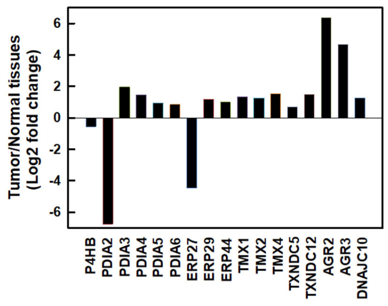
We first investigated messenger (m)RNA expressions of PDI family members using the Gene Expression Profiling Interactive Analysis 2 (GEPIA2) database [25] in normal human pancreatic and PDAC tissues. As shown in Figure 1, most PDI family members were upregulated except for PDIA2 and ERP27 in human PDAC tissues. Compared to normal pancreatic tissues, the top three members with the highest fold changes in expression in human PDAC tissues were AGR2 (fold change 6.392), AGR3 (fold change 4.689), and PDIA3 (fold change 1.961). These results suggested that PDI family members might play important roles in PDAC cell proliferation.
我们首先使用基因表达分析交互式分析 2 (GEPIA2) 数据库 [ 25 ] 在正常人胰腺和 PDAC 组织中研究了 PDI 家族成员的信使 (m)RNA 表达。如图所示 Figure 1 ,除人 PDAC 组织中 PDIA2 和 ERP27 外,大多数 PDI 家族成员均上调。与正常胰腺组织相比,人 PDAC 组织中表达倍数变化最高的前三个成员是 AGR2 (倍数变化 6.392) 、 AGR3 (倍数变化 4.689) 和 PDIA3 (倍数变化 1.961)。这些结果表明 PDI 家族成员可能在 PDAC 细胞增殖中发挥重要作用。
Figure 1. 图 1.
The mRNA expressions of protein disulfide isomerase (PDI) family members in human pancreatic ductal adenocarcinoma (PDAC) tissues. mRNA levels of human PDAC tissues and normal pancreatic tissues were obtained from the GEPIA2 database. PDI family members were selected based on a |log2(fold change)| cutoff of 0.5 and q-value cutoff of 0.01. PDAC tissues, n = 179; normal pancreatic tissues, n = 171.
人胰腺导管腺癌 (PDAC) 组织中蛋白二硫键异构酶 (PDI) 家族成员的 mRNA 表达。人 PDAC 组织和正常胰腺组织的 mRNA 水平从 GEPIA2 数据库获得。PDI 家族成员是根据 |log2(fold change)|截止值为 0.5,Q 值截止值为 0.01。PDAC 组织,n = 179;正常胰腺组织,n = 171。
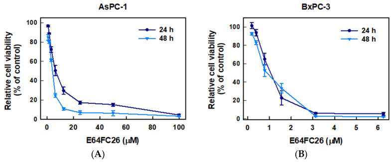
To examine whether inhibition of PDI activity could decrease the proliferation of PDAC cells, we used E64FC26, a novel PDI inhibitor, to treat human pancreatic cancer cell lines AsPC-1 and BxPC-3 cells for 24 and 48 h and determined cell viability by an MTT assay. As shown in Figure 2, E64FC26 significantly decreased the viability of both cell lines in dose-dependent manners. The 50% inhibitory concentrations (IC50) with 24 h of treatment were 6.13 ± 0.08 and 0.93 ± 0.33 µM in AsPC-1 and BxPC-3 cells, respectively; and IC50 values after 48 h of treatment were 3.41 ± 0.11 and 0.87 ± 0.16 µM, respectively. Prolonged drug treatment for 48 h was more effective than 24 h treatment in AsPC-1 cells, but there was no significant difference in BxPC-3 cells. These results suggest that the E64FC26 PDI inhibitor significantly inhibited proliferation of PDAC cells.
为了检查抑制 PDI 活性是否可以减少 PDAC 细胞的增殖,我们使用新型 PDI 抑制剂 E64FC26 处理人胰腺癌细胞系 AsPC-1 和 BxPC-3 细胞 24 和 48 小时,并通过 MTT 测定测定细胞活力。如 Figure 2 所示,E64FC26 以剂量依赖性方式显著降低两种细胞系的活力。AsPC-1 和 BxPC-3 细胞处理 24 h 的 50% 抑制浓度 (IC50) 分别为 6.13 ± 0.08 和 0.93 ± 0.33 μM;处理 48 小时后的 IC50 值分别为 3.41 ± 0.11 和 0.87 ± 0.16 μM。在 AsPC-1 细胞中,延长药物治疗 48 h 比处理 24 h 更有效,但在 BxPC-3 细胞中无显著差异。这些结果表明,E64FC26 PDI 抑制剂显着抑制了 PDAC 细胞的增殖。
Figure 2. 图 2.
Effects of E64FC26 on the cell viability in human pancreatic ductal adenocarcinoma (PDAC) cells. (A) AsPC-1 and (B) BxPC-3 cells were treated with different concentrations of E64FC26 for 24 and 48 h, and cell viability was determined by an MTT assay. Each data point is presented as the mean ± S.E. of three independent experiments.
E64FC26 对人胰腺导管腺癌 (PDAC) 细胞活力的影响。(A) AsPC-1 和 (B) BxPC-3 细胞用不同浓度的 E64FC26 处理 24 和 48 h,并通过 MTT 测定细胞活力。每个数据点都表示为三个独立实验的均值± SE。
2.2. The PDI Inhibitor, E64FC26, Induced ER Stress and the UPR
2.2. PDI 抑制剂 E64FC26 诱导的 ER 应激和 UPR
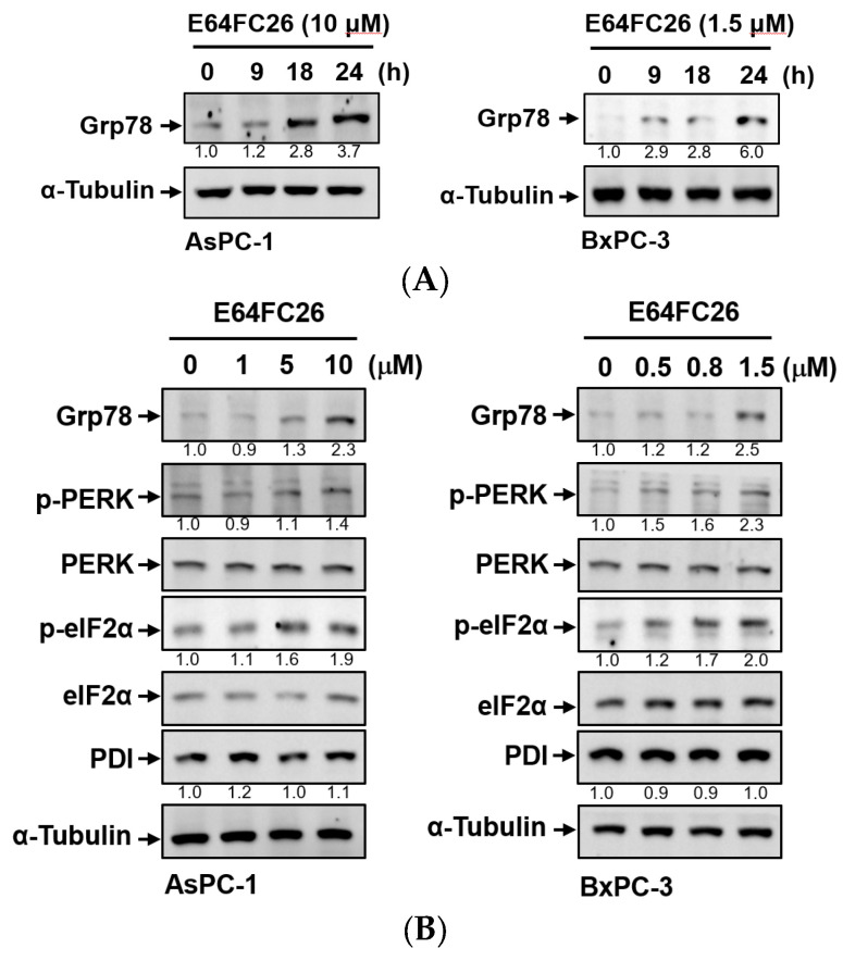
PDI inhibitors are known to be able to impede the correct disulfide bond formation of nascent proteins and cause protein misfolding [26]. Accumulation of misfolded proteins can increase ER stress and the UPR. To explore whether E64FC26 could induce ER stress and the UPR in PDAC cells, we detected protein expressions of ER stress- and UPR-related signaling pathways. Grp78, a major ER stress-inducible chaperone, was time-dependently upregulated by E64FC26 treatment in both cell lines (Figure 3A). E64FC26 also significantly increased Grp78 expressions in dose-dependent manners in both cell lines (Figure 3B). E64FC26-induced ER stress led to significant PERK phosphorylation, and activated PERK then directly phosphorylated eIF2α in both cell lines. E64FC26 did not change the PDI protein expression in either cell line (Figure 3B). These results suggest that the PDI inhibitor, E64FC26, significantly increased ER stress and the UPR in PDAC cells.
已知 PDI 抑制剂能够阻碍新生蛋白质的正确二硫键形成并导致蛋白质错误折叠 [ 26 ]。错误折叠蛋白的积累会增加 ER 应激和 UPR。为了探讨 E64FC26 是否可以诱导 PDAC 细胞中的 ER 应激和 UPR,我们检测了 ER 应激和 UPR 相关信号通路的蛋白表达。Grp78 是一种主要的 ER 应激诱导伴侣,在两种细胞系中,E64FC26处理均呈时间依赖性上调 ( Figure 3 A)。E64FC26 还以剂量依赖性方式显着增加了两种细胞系中 Grp78 的表达 ( Figure 3 B)。E64FC26诱导的 ER 应激导致显著的 PERK 磷酸化,激活的 PERK 随后在两种细胞系中直接磷酸化 eIF2α。E64FC26 没有改变两种细胞系 ( Figure 3 B) 中的 PDI 蛋白表达。这些结果表明,PDI 抑制剂 E64FC26 显著增加了 PDAC 细胞的 ER 应激和 UPR。
Figure 3. 图 3.
Effects of E64FC26 on endoplasmic reticular (ER) stress- and unfolded protein response (UPR)-related protein expressions in human pancreatic ductal adenocarcinoma (PDAC) cells. (A) AsPC-1 and BxPC-3 cells were treated with E64FC26 for different times, and protein levels of ER stress-related proteins were determined by Western blotting. (B) AsPC-1 and BxPC-3 cells were treated with different concentrations of E64FC26 for 24 h, and protein levels of ER stress-related proteins were determined by Western blotting. The relative mean intensity of each band (indicated below the bands, n ≥ 3) was normalized to the unphosphorylated total protein (including PERK, eIF2α) or α-tubulin loading control.
E64FC26 对人胰腺导管腺癌 (PDAC) 细胞中内质网 (ER) 应激和未折叠蛋白反应 (UPR) 相关蛋白表达的影响。(A) AsPC-1 和 BxPC-3 细胞用 E64FC26 处理不同时间,通过 Western blotting 测定 ER 应激相关蛋白的蛋白水平。(B) 用不同浓度的 E64FC26 处理 AsPC-1 和 BxPC-3 细胞 24 h,通过 Western blotting 测定 ER 应激相关蛋白的蛋白水平。将每个条带的相对平均强度(显示在条带下方,n ≥ 3)归一化为未磷酸化的总蛋白(包括 PERK,eIF2α)或 α-微管蛋白负载对照。
2.3. The PDI Inhibitor, E64FC26, Caused Cell Death Partially via Apoptosis
2.3. PDI 抑制剂 E64FC26 部分通过细胞凋亡导致细胞死亡
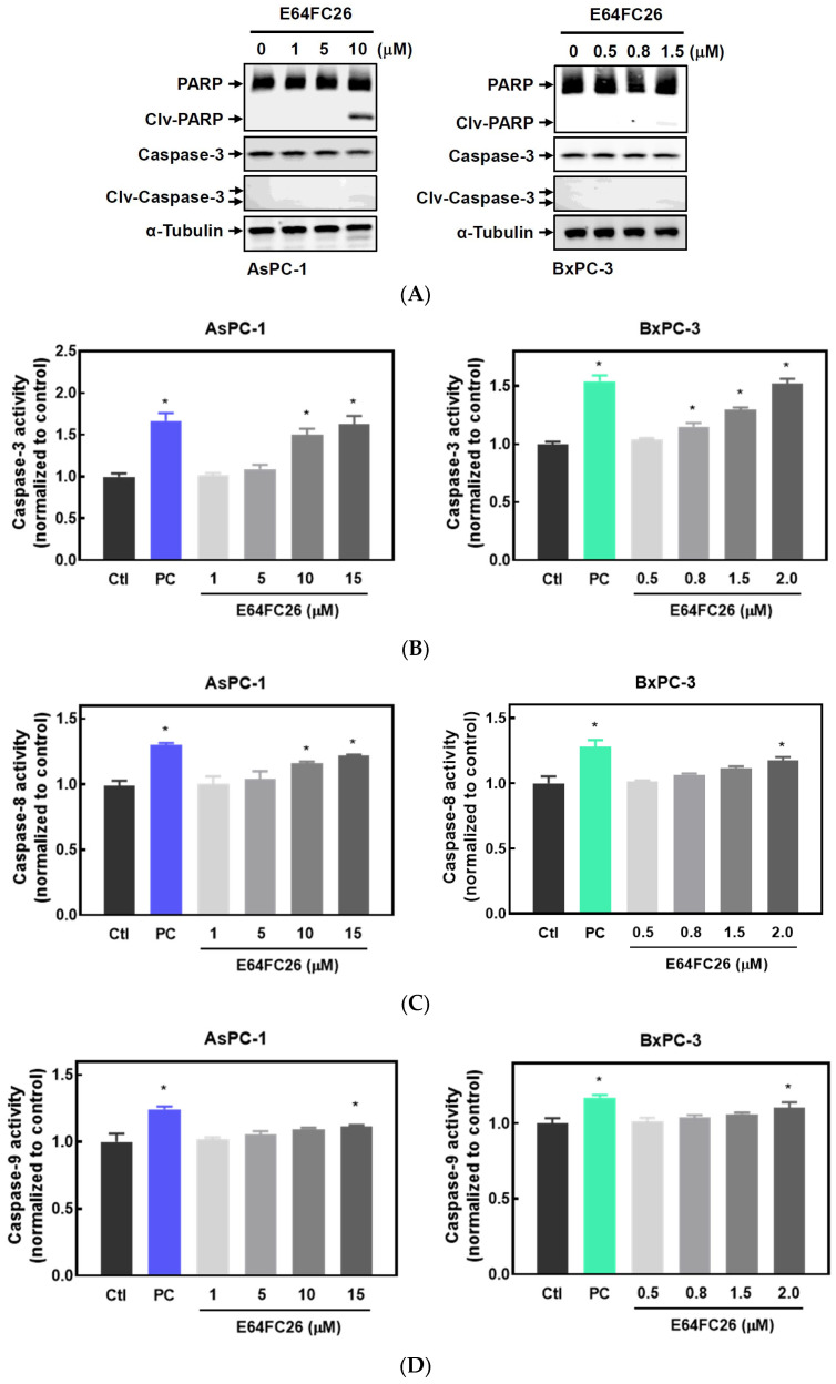
It is known that persistent ER stress and the UPR can eventually trigger cell apoptosis [27]. To examine whether the PDI inhibitor, E64FC26, can induce apoptosis in PDAC cells, we detected expressions of cleaved caspases by Western blotting and caspase activities. E64FC26 at 10 and 1.5 µM in AsPC-1 and BxPC-3 cells, respectively, induced very limited expressions of cleaved PARP and cleaved caspase-3 (Figure 4A), but cleaved caspase-8 and caspase-9 were absent from Western blots. To further confirm whether E64FC26 can induce cell apoptosis in PDAC cells, we treated cells with a higher concentration of E64FC26 and directly detected caspase activity with a commercially available caspase activity assay kit. After increasing the E64FC26 concentration to 15 and 2 µM, respectively, in AsPC-1 and BxPC-3 cells, E64FC26 significantly induced caspase-3, -8, and -9 activities using a caspase activity assay kit (Figure 4B). These results suggest that the PDI inhibitor, E64FC26, partially induced cell death through apoptosis in PDAC cells.
众所周知,持续的 ER 应激和 UPR 最终会触发细胞凋亡 [ 27 ]。为了检查 PDI 抑制剂 E64FC26 是否可以诱导 PDAC 细胞凋亡,我们通过 Western blotting 和 caspase 活性检测了裂解的 caspase 的表达。在 AsPC-1 和 BxPC-3 细胞中分别以 10 μM 和 1.5 μM E64FC26诱导非常有限的裂解 PARP 和裂解的 caspase-3 ( Figure 4 A) 的表达,但裂解的 caspase-8 和 caspase-9 在蛋白质印迹中不存在。为了进一步确认 E64FC26 是否可以诱导 PDAC 细胞凋亡,我们用较高浓度的 E64FC26 处理细胞,并用市售的 caspase 活性测定试剂盒直接检测 caspase 活性。在 AsPC-1 和 BxPC-3 细胞中将 E64FC26 浓度分别增加到 15 和 2 μM 后,E64FC26 使用 caspase 活性测定试剂盒 ( Figure 4 B) 显著诱导 caspase-3 、 -8 和 -9 活性。这些结果表明,PDI 抑制剂 E64FC26 通过 PDAC 细胞凋亡部分诱导细胞死亡。
Figure 4. 图 4.
Effects of E64FC26 on activation of caspases in human pancreatic ductal adenocarcinoma (PDAC) cells. (A) AsPC-1 and BxPC-3 cells were treated with different concentrations of E64FC26 for 24 h, and protein levels of the poly(ADP ribose) polymerase (PARP) and caspase-3 proteins were determined by Western blotting. (B–D) AsPC-1 and BxPC-3 cells were treated with different concentrations of E64FC26 for 24 h, and activities of (B) caspase-3, (C) caspase-8, and (D) caspase-9 were detected with a caspase colorimetric assay kit. Concentrations of 1 µM and 100 nM of staurosporine were respectively used as positive controls (PCs) in AsPC-1 and BxPC-3 cells. Each data point is presented as the mean ± S.E. of three independent experiments. * p < 0.05 vs. the control (Ctl) group.
E64FC26 对人胰腺导管腺癌 (PDAC) 细胞中 caspase 活化的影响。(A) 用不同浓度的 E64FC26 处理 AsPC-1 和 BxPC-3 细胞 24 h,Western blotting 测定聚 (ADP 核糖) 聚合酶 (PARP) 和 caspase-3 蛋白的蛋白水平。(B-D)用不同浓度的 E64FC26 处理 AsPC-1 和 BxPC-3 细胞 24 h,用 caspase 比色测定试剂盒检测 (B) caspase-3 、 (C) caspase-8 和 (D) caspase-9 的活性。浓度为 1 μM 和 100 nM 的星形孢菌素分别用作 AsPC-1 和 BxPC-3 细胞中的阳性对照 (PC)。每个数据点都表示为三个独立实验的均值± SE。* p < 0.05 vs. 对照 (Ctl) 组。
2.4. PDI Inhibitor E64FC26-Induced Cell Death Might Be Associated with Ferroptosis
2.4. PDI 抑制剂 E64FC26 诱导的细胞死亡可能与铁死亡有关
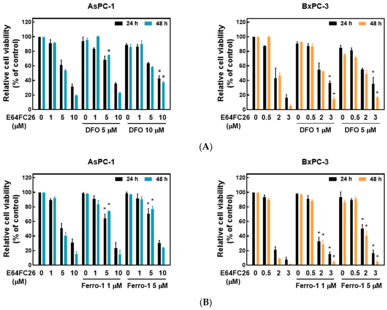
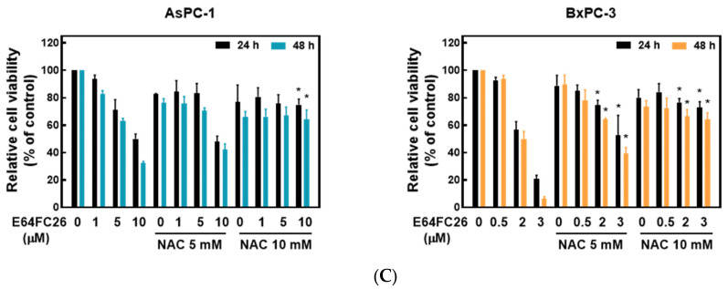
Ferroptosis is a kind of non-apoptotic cell death that depends on iron and lipid peroxidation (LPO) [28]. It was reported that upregulation of ER stress contributes to ferroptosis [29]. To investigate whether E64FC26 caused cell death through ferroptosis, we used the iron-chelating agent, deferoxamine (DFO), and the ROS scavengers, ferrostatin-1 and NAC, to reverse E64FC26-caused cell death. As shown in Figure 5A, pretreatment with 5 and 10 µM of DFO significantly reversed 5 and 10 µM of E64FC26-induced cell death, respectively, in AsPC-1 cells with both 24 and 48 h of treatment. Although DFO alone caused partial cell death, 1 and 5 µM of DFO also reversed 3 µM E64FC26-induced cell death in BxPC-3 cells at both 24 and 48 h of treatment. At both 24 and 48 h, 1 and 5 µM of ferrostatin-1 reversed the 5 µM E64FC26-induced death of AsPC-1 cells, and 2 and 3 µM of E64FC26-induced cell death in BxPC-3 cells (Figure 5B). In addition, 10 mM of NAC prevented cell death under 10 µM E64FC26 treatment for both 24 and 48 h in AsPC-1 cells, and 5 and 10 mM of NAC increased cell survival under 2 and 3 µM of E64FC26 treatment at both 24 and 48 h in BxPC-3 cells (Figure 5C). Interestingly, we detected no significant increase in ROS by the cytosolic ROS sensor, 2’-7’-dichlorodihydrofluorescein diacetate (DCF-DA), in E64FC26-treated cells. These results suggest that E64FC26-caused cell death might be partially mediated through ferroptosis in PDAC cells.
铁死亡是一种非凋亡性细胞死亡,依赖于铁和脂质过氧化 (LPO) [ 28 ]。据报道,ER 应激的上调会导致铁死亡 [ 29 ]。为了研究 E64FC26 是否通过铁死亡导致细胞死亡,我们使用铁螯合剂去铁胺 (DFO) 和 ROS 清除剂铁司他汀-1 和 NAC 来逆转 E64FC26 引起的细胞死亡。如 Figure 5 A 所示,在处理 24 小时和 48 小时的 AsPC-1 细胞中,用 5 μM 和 10 μM DFO 预处理分别显着逆转了 5 μM 和 10 μM E64FC26诱导的细胞死亡。尽管单独的 DFO 导致部分细胞死亡,但在处理的 24 小时和 48 小时,1 μM 和 5 μM 的 DFO 也逆转了 BxPC-3 细胞中 3 μM E64FC26诱导的细胞死亡。在 24 和 48 小时,1 μM 和 5 μM 的 ferrostatin-1 逆转了 5 μM E64FC26诱导的 AsPC-1 细胞死亡,以及 BxPC-3 细胞中 2 μM 和 3 μM E64FC26诱导的细胞死亡 ( Figure 5 B)。此外,在 AsPC-1 细胞中,10 mM 的 NAC 在 10 μM E64FC26处理下 24 和 48 小时可防止细胞死亡,并且在 2 μM 和 3 μM E64FC26处理下,5 和 10 mM 的 NAC 在 BxPC-3 细胞中 24 和 48 小时均能增加细胞存活率 ( Figure 5 C)。有趣的是,我们在 E64FC26 处理的细胞中检测到胞质 ROS 传感器 2'-7'-二氯二氢荧光素二乙酸酯 (DCF-DA) 的 ROS 没有显着增加。这些结果表明,E64FC26 引起的细胞死亡可能是通过 PDAC 细胞中的铁死亡部分介导的。
Figure 5. 图 5.
Effects of ferroptosis inhibitors on E64FC26-caused cell death in human pancreatic ductal adenocarcinoma (PDAC) cells. AsPC-1 and BcPC-3 cells were pretreated with (A) deferoxamine (DFO) for 6 h, (B) ferrostatin-1 (Ferro-1) for 1 h, or (C) N-acetylcysteine (NAC) for 24 h, and then treated with E64FC26 for 24 or 48 h. Cell viability was determined by an MTT assay. Each data point is presented as the mean ± S.E. of three independent experiments. * p < 0.05 vs. the E64FC26 alone group.
铁死亡抑制剂对人胰腺导管腺癌 (PDAC) 细胞中 E64FC26 引起的细胞死亡的影响。AsPC-1 和 BcPC-3 细胞用 (A) 去铁胺 (DFO) 预处理 6 h,(B) ferrostatin-1 (Ferro-1) 预处理 1 h,或 (C) N-乙酰半胱氨酸 (NAC) 预处理 24 h,然后用 E64FC26 处理 24 或 48 h。通过 MTT 测定法测定细胞活力。每个数据点都表示为三个独立实验的均值± SE。* p < 0.05 vs. 单独E64FC26组。
2.5. The PDI Inhibitor, E64FC26, Caused Autophagic Cell Death by Blocking Autolysosome Formation
2.5. PDI 抑制剂 E64FC26 通过阻断自噬溶酶体的形成导致自噬细胞死亡
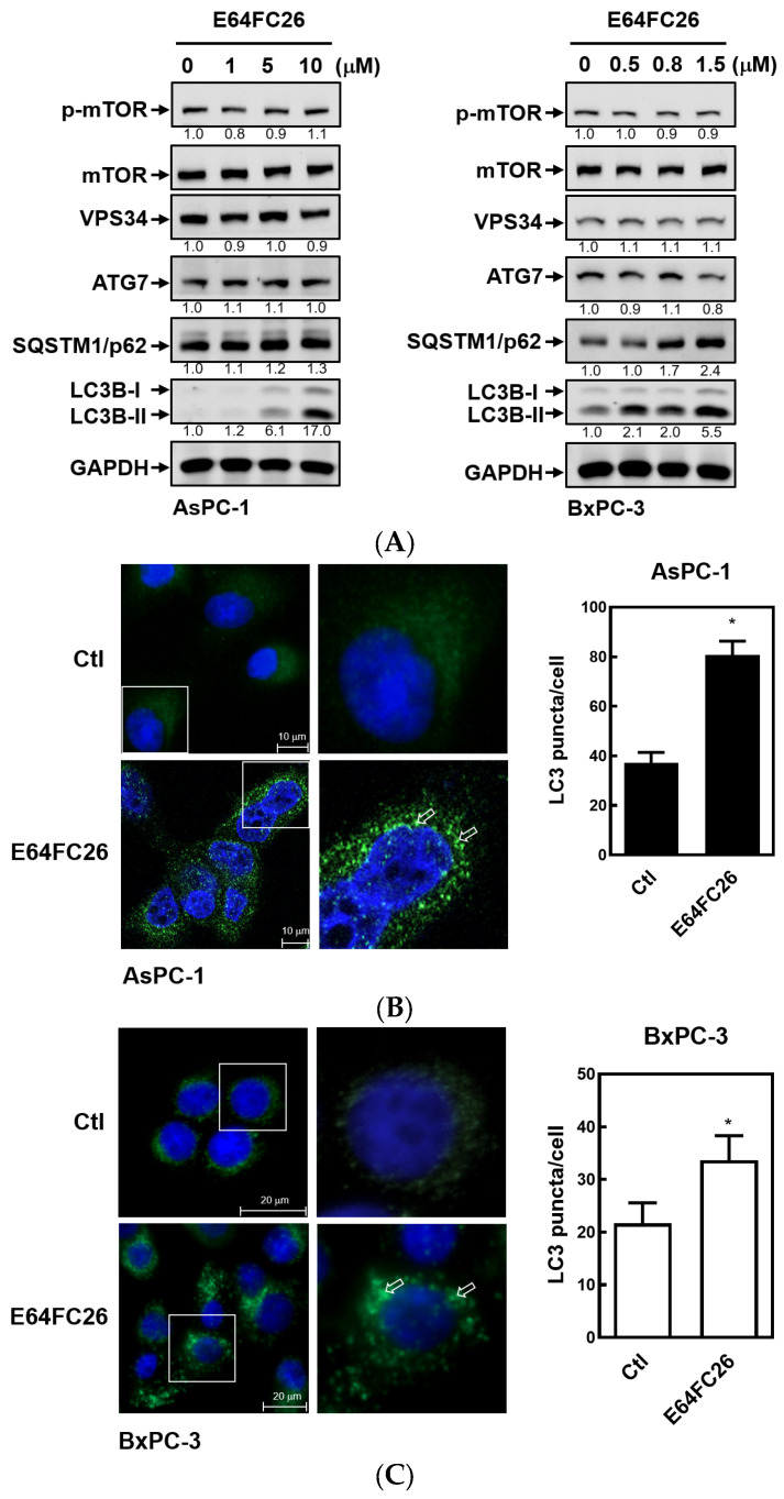
Previous studies demonstrated that PDAC cells have a high basal level of autophagy, which enables them to continue cell proliferation in vivo and in vitro and enhances resistance to chemotherapy and radiation therapy [30,31]. LC3B-II is commonly used as an indicator of mammalian autophagosome formation. To further examine whether the PDI inhibitor, E64FC26, can induce cell death through autophagy, we detected LC3B-II expression by Western blotting and LC3B puncta formation in PDAC cells. As shown in Figure 6A, E64FC26 treatment resulted in LC3B-II accumulation in a dose-dependent manner in both cell lines. Moreover, E64FC26 increased LC3B puncta formation as revealed by IF staining (Figure 6B,C). Expressions of components of autophagy initiation, including VPS34 and Atg7, did not change upon E64FC26 treatment. The final process of autophagy is the fusion of autophagosomes and lysosomes into autolysosomes, which allows degradation of their contents, including SQSTM1/p62. Interestingly, E64FC26 did not induce SQSTM1/p62 degradation but significantly increased SQSTM1/p62 expressions in a dose-dependent manner in both cell lines (Figure 6A). These results suggest that E64FC26 induced incomplete autophagy by increasing the LC3B-II level but failed to degrade SQSTM1/p62.
先前的研究表明,PDAC 细胞具有高水平的基础自噬,这使它们能够在体内和体外继续细胞增殖,并增强对化疗和放疗的抵抗力 [ 30 , 31 ]。LC3B-II 通常用作哺乳动物自噬体形成的指标。为了进一步研究 PDI 抑制剂 E64FC26 是否可以通过自噬诱导细胞死亡,我们通过 Western blotting 检测 LC3B-II 在 PDAC 细胞中的表达和 LC3B 点状形成。如 Figure 6 A 所示,E64FC26处理导致 LC3B-II 在两种细胞系中以剂量依赖性方式积累。此外,E64FC26 增加了 IF 染色 ( Figure 6 B,C) 显示的 LC3B 点形成。自噬起始成分的表达,包括 VPS34 和 Atg7,在 E64FC26 处理后没有变化。自噬的最后一个过程是自噬体和溶酶体融合成自噬溶酶体,这允许降解其内容物,包括 SQSTM1/p62。有趣的是,E64FC26 没有诱导 SQSTM1/p62 降解,但在两种细胞系中以剂量依赖性方式显着增加 SQSTM1/p62 表达 ( Figure 6 A)。这些结果表明,E64FC26通过增加 LC3B-II 水平诱导不完全自噬,但未能降解 SQSTM1/p62。
Figure 6. 图 6.
Effects of E64FC26 on autophagy marker protein expressions in human pancreatic ductal adenocarcinoma (PDAC) cells. (A) AsPC-1 and BxPC-3 cells were treated with different concentrations of E64FC26 for 24 h, and protein levels of autophagy-related proteins were determined by Western blotting. The relative mean intensity of each band (indicated below the bands, n ≥ 3) was normalized to the unphosphorylated total protein (mTOR) or GAPDH loading control. (B) AsPC-1 and (C) BxPC-3 cells were respectively treated with 5 and 1 µM of E64FC26 for 24 h, and the light chain 3B (LC3B) puncta were detected by IF staining with the CF®488A dye (green) and nucleic acid staining with DAPI (blue). Images on the right column are magnifications of the white boxed areas. Representative LC3B puncta are indicated by white arrows. Quantification of LC3 puncta per cell is presented as the mean ± S.E. of three independent experiments. * p < 0.05 vs. the control (Ctl) group.
E64FC26 对人胰腺导管腺癌 (PDAC) 细胞自噬标志物蛋白表达的影响。(A) 用不同浓度的 AsPC-1 和 BxPC-3 细胞E64FC26处理 24 h,Western blotting 测定自噬相关蛋白的蛋白水平。将每个条带的相对平均强度(显示在条带下方,n ≥ 3)归一化为未磷酸化总蛋白 (mTOR) 或 GAPDH 上样对照。(B) AsPC-1 和 (C) BxPC-3 细胞分别用 5 μM 和 1 μM E64FC26处理 24 h,通过 CF®488A 染料 (绿色) IF 染色和 DAPI (蓝色) 核酸染色检测轻链 3B (LC3B) 点。右列的图像是白色框区域的放大倍数。代表性的 LC3B 点状物用白色箭头表示。每个细胞 LC3 点的定量表示为三个独立实验的平均 ± SE。* p < 0.05 vs. 对照 (Ctl) 组。
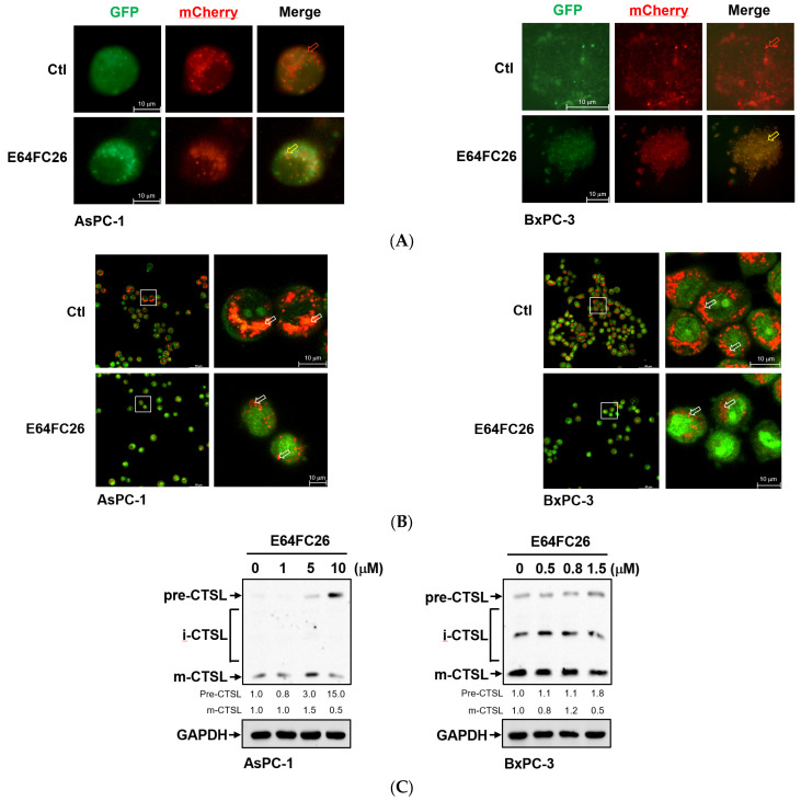
To investigate why SQSTM1/p62 was not degraded with E64FC26 treatment, we first transfected the autophagy reporter plasmid FUW mCherry-GFP-LC3 into PDAC cells to express the mCherry-GFP-LC3 fusion protein. The mCherry protein is stable at a neutral pH and in an acidic condition, while the GFP protein is only stable at neutral pH but is acid-labile. Therefore, the mCherry-GFP-LC3 fusion protein displays both green and red fluorescence in the neutral environment of the autophagosome lumen, whereas it only exhibits red fluorescence in the acidic condition of the autolysosome lumen. Compared to control cells, E64FC26 treatment significantly increased yellow fluorescent spots in both cell lines (Figure 7A), indicating that both mCherry and GFP were expressed in autophagosomes of these cells. These results suggest that E64FC26 treatment might block autolysosome formation through inhibiting the fusion of autophagosomes and lysosomes in PDAC cells. To further confirm whether E64FC26 blocks the fusion of autophagosomes and lysosomes, we used AO dye to stain acidic vesicles of cells. As shown in Figure 7B, control cells displayed a lot of reddish-orange fluorescence dots, indicating the formation of mature lysosomes and acidic autolysosomes. However, cells treated with E64FC26 exhibited few reddish-orange fluorescence dots, indicating that E64FC26 blocked autolysosome formation or induced lysosome defects. CTSL is a lysosomal protease that is activated by lysosomal cleavage of pre-CTSL to form mature CTSL. To examine whether E64FC26 impaired lysosome function, we detected expressions of pre-CTSL and mature CTSL by Western blotting in PDAC cells. As shown in Figure 7C, E64FC26 treatment dose-dependently caused the accumulation of pre-CTSL, but decreased mature CTSL in both cell lines. These results suggest that E64FC26 might block autolysosome formation through inducing lysosome defects in PDAC cells.
为了研究 SQSTM1/p62 在 E64FC26 处理后未降解的原因,我们首先将自噬报告基因质粒 FUW mCherry-GFP-LC3 转染到 PDAC 细胞中以表达 mCherry-GFP-LC3 融合蛋白。mCherry 蛋白在中性 pH 值和酸性条件下稳定,而 GFP 蛋白仅在中性 pH 值下稳定,但耐酸性。因此,mCherry-GFP-LC3 融合蛋白在自噬体管腔的中性环境中同时显示绿色和红色荧光,而它仅在自噬溶酶体管腔的酸性条件下显示红色荧光。与对照细胞相比,E64FC26处理显着增加了两种细胞系 ( Figure 7 A) 中的黄色荧光斑点,表明 mCherry 和 GFP 在这些细胞的自噬体中均表达。这些结果表明,E64FC26治疗可能通过抑制 PDAC 细胞中自噬体和溶酶体的融合来阻止自噬溶酶体的形成。为了进一步确认 E64FC26 是否阻断自噬体和溶酶体的融合,我们使用 AO 染料对细胞的酸性囊泡进行染色。如 Figure 7 B 所示,对照细胞显示大量红橙色荧光点,表明成熟溶酶体和酸性自溶酶体的形成。然而,用 E64FC26 处理的细胞表现出很少的红橙色荧光点,表明E64FC26阻断了自噬溶酶体的形成或诱导了溶酶体缺陷。CTSL 是一种溶酶体蛋白酶,可被 pre-CTSL 的溶酶体裂解激活以形成成熟的 CTSL。为了检查E64FC26溶酶体功能是否受损,我们通过 Western blotting 检测 PDAC 细胞中 pre-CTSL 和成熟 CTSL 的表达。 如 Figure 7 C 所示,E64FC26 处理剂量依赖性地导致 pre-CTSL 的积累,但在两种细胞系中成熟 CTSL 降低。这些结果表明,E64FC26 可能通过诱导 PDAC 细胞中的溶酶体缺陷来阻断自体溶酶体的形成。
Figure 7. 图 7.
Effects of E64FC26 on autolysosome formation and lysosome functions in human pancreatic ductal adenocarcinoma (PDAC) cells. (A) AsPC-1 and BxPC-3 cells were transfected with the FUW mCherry-GFP-LC3 plasmid and then treated with 5 µM and 1 µM E64FC26, respectively, for 24 h. Autophagosomes/autolysosomes were visualized by fluorescence microscopy. Representative autolysosomes and autophagosomes are indicated by red and yellow arrows, respectively. (B) AsPC-1 and BxPC-3 cells were treated with 5 µM and 1 µM E64FC26, respectively, for 24 h, and cells were stained with acridine orange for 15 min. Under acridine orange staining, cytoplasmic and nuclear fluorescence was green, and acidic vesicular organelle fluorescence was bright red or orange-red. Images on the right column are magnifications of the white boxed areas. Representative mature lysosomes or acidic autolysosomes are indicated by white arrows. (C) AsPC-1 and BxPC-3 cells were treated with different concentrations of E64FC26 for 24 h, and the precursor (pre-), intermediate (i-), and mature (m-) forms of the cathepsin L (CTSL) protein were detected by Western blotting. The relative mean intensity of each band (indicated below the bands, n ≥ 3) was normalized to the GAPDH loading control.
E64FC26 对人胰腺导管腺癌 (PDAC) 细胞中自体溶酶体形成和溶酶体功能的影响。(A) 用 FUW mCherry-GFP-LC3 质粒转染 AsPC-1 和 BxPC-3 细胞,然后分别用 5 μM 和 1 μM E64FC26处理 24 h。自噬体/自噬溶酶体通过荧光显微镜观察。代表性自噬溶酶体和自噬体分别用红色和黄色箭头表示。(B) AsPC-1 和 BxPC-3 细胞分别用 5 μM 和 1 μM E64FC26处理 24 h,并用吖啶橙染色 15 min。在吖啶橙染色下,细胞质和细胞核荧光为绿色,酸性囊泡细胞器荧光为亮红色或橙红色。右列的图像是白色框区域的放大倍数。代表性成熟溶酶体或酸性自溶酶体用白色箭头表示。(C) 用不同浓度的 E64FC26 处理 AsPC-1 和 BxPC-3 细胞 24 h,Western blotting 检测组织蛋白酶 L (CTSL) 蛋白的前体 (pre-)、中间体 (i-) 和成熟 (m-) 形式。将每个条带的相对平均强度(显示在条带下方,n ≥ 3)归一化为 GAPDH 负载对照。
3. Discussion 3. 讨论
At least 21 members of the human PDI family have been discovered. The main functions of PDI members include assisting the formation of disulfide bonds in nascent proteins and serving as chaperones to assist the correct folding of proteins. Actively proliferating tumor cells require correct protein folding, and many PDI members are therefore often highly expressed in tumor cells. The development of PDI inhibitors may be a new strategy to fight tumors [20], although no PDI inhibitors have yet entered clinical trials or become Food and Drug Administration (FDA)-approved drugs. Exploring the underlying molecular mechanisms of PDI inhibitors will improve the development of PDI drugs and the feasibility of combining them with other drug treatments. Several PDI members, including PDIA3, PDIA4, PDIA6, ERP29, and TXNDC5, were found to be highly expressed in numerous cancer types [32]. In addition to those PDI members, PDIA5, ERP44, TMX1, TMX2, TMX4, TXNDC12, AGR2, AGR3, and DNAJC10 were also determined to be upregulated in PDAC (Figure 1). It is worth noting that PDIA1, PDIA2, and ERP27 are highly expressed in the normal pancreas but are significantly downregulated in PDAC. In this study, we focused on the recently developed novel PDI inhibitor, E64FC26, for treating refractory pancreatic cancer. We found that E64FC26 might disrupt proteostasis and lead to cell death through multiple pathways in PDAC cells. E64FC26 might be developed as a potent anti-PDAC drug either alone or in combination with other anticancer drugs.
已发现至少 21 名人类 PDI 家族成员。PDI 成员的主要功能包括协助新生蛋白质中形成二硫键,并作为伴侣协助蛋白质的正确折叠。活跃增殖的肿瘤细胞需要正确的蛋白质折叠,因此许多 PDI 成员通常在肿瘤细胞中高度表达。PDI 抑制剂的开发可能是对抗肿瘤的一种新策略 [ 20 ],尽管尚未有 PDI 抑制剂进入临床试验或成为美国食品和药物管理局 (FDA) 批准的药物。探索 PDI 抑制剂的潜在分子机制将改善 PDI 药物的开发以及将其与其他药物治疗联合使用的可行性。发现几种 PDI 成员,包括 PDIA3、PDIA4、PDIA6、ERP29 和 TXNDC5,在许多癌症类型中高度表达 [ 32 ]。除了这些 PDI 成员之外,PDIA5、ERP44、TMX1、TMX2、TMX4、TXNDC12、AGR2、AGR3 和 DNAJC10 也被确定在 PDAC 中上调 ( Figure 1 )。值得注意的是,PDIA1 、 PDIA2 和 ERP27 在正常胰腺中高度表达,但在 PDAC 中显著下调。在这项研究中,我们专注于最近开发的新型 PDI 抑制剂 E64FC26,用于治疗难治性胰腺癌。我们发现E64FC26可能会破坏蛋白质稳态并通过 PDAC 细胞中的多种途径导致细胞死亡。E64FC26 可能单独开发为有效的抗 PDAC 药物,也可以与其他抗癌药物联合开发。
Previous studies have found that several reversible inhibitors bind to PDI and inhibits their activities, including 35G8 [24], bepristat 2a [15], ML359 [33], LOC14 [34], and rutin [35]. On the other hand, many covalent inhibitors of PDI have been discovered in the past few years, such as 16F16 [11], CCF642 [21], PACMA31 [19], KSC-34 [36], and E64FC26. In general, reversible inhibitors of PDI are less effective than covalently bound PDI inhibitors, and there is a trend in the number of drugs using covalent inhibitors to treat cancer patients. However, whether covalent inhibitors of PDI can specifically bind to PDI but not other molecules containing SH groups is an important question. The catechol moiety of E64FC26 [(E)-1-nonylidene-3-(trifluoromethyl)-1H-indene-5,6-diol] can be oxidized intracellularly into the o-quinone form, which is further reacts with thiol-containing molecules, such as cysteine residue of proteins and glutathione, to form covalent bonds [37]. The electrophilic trifluoro group of E64FC26 was designed for attaching the nucleophilic cysteines of the PDI active site, but other trifluoro-containing molecules could not become effective PDI inhibitors [38]. Therefore, E64FC26 is considered a PDI inhibitor with higher specificity. E64FC26 was reported to be a potent pan-PDI inhibitor, and is more effective against PDIA1, PDIA3, PDIA4, PDIA6, and TXNDC5 [22]. However, the characteristic of E64FC26 reacting with thiol groups cannot rule out its reaction with non-PDI molecules, especially when body fluids contain very high concentrations of glutathione.
以前的研究发现,几种可逆抑制剂与 PDI 结合并抑制其活性,包括 35G8 [ 24 ]、bepristat 2a [ 15 ]、ML359 [ 33 ]、LOC14 [ 34 ] 和芦丁 [ 35 ]。另一方面,在过去几年中发现了许多 PDI 的共价抑制剂,例如 16F16 [ 11 ]、CCF642 [ 21 ]、PACMA31 [ 19 ]、KSC-34 [ 36 ] 和 E64FC26。一般来说,PDI 的可逆抑制剂不如共价结合的 PDI 抑制剂有效,并且使用共价抑制剂治疗癌症患者的药物数量呈趋势。然而,PDI 的共价抑制剂是否可以特异性结合 PDI,但不能特异性结合其他含有 SH 基团的分子是一个重要的问题。E64FC26 [(E)-1-壬二烯-3-(三氟甲基)-1H-茚-5,6-二醇] 的儿茶酚部分可在细胞内氧化成邻醌形式,进一步与含巯基的分子反应,如蛋白质的半胱氨酸残基和谷胱甘肽,形成共价键 [ 37 ]。E64FC26的亲电三氟基团被设计为连接 PDI 活性位点的亲核半胱氨酸,但其他含三氟的分子不能成为有效的 PDI 抑制剂 [ 38 ]。因此,E64FC26 被认为是具有更高特异性的 PDI 抑制剂。据报道,E64FC26 是一种有效的泛 PDI 抑制剂,对 PDIA1、PDIA3、PDIA4、PDIA6 和 TXNDC5 更有效 [ 22 ]。然而,E64FC26与硫醇基反应的特性不能排除它与非 PDI 分子的反应,尤其是当体液中含有非常高浓度的谷胱甘肽时。
Activating mutations in oncogenic KRAS are very common in pancreatic cancer, where they are persistently activated and induce multiple proliferative signaling pathways. KRAS mutations were found in AsPC-1 cells, while BxPC-3 cells were identified as wild type [39]. Adhesion ability is important for cell migration and affects cell proliferation and invasion in vivo and in vitro [40]. The adherent potential and metastatic activity of BxPC-3 cells were lower than those of AsPC-1 cells [39]. Compared with AsPC-1 cells, lower adhesion and metastasis abilities as well as wild-type KRAS might cause BxPC-3 cells to be more toxic to E64FC26 treatment. As previous studies have demonstrated, gene mutation, such as KRAS, EMT phenotype, and cell–cell adhesion could affect drug sensitivity in PDAC cells [41]. AsPC-1 is generally regarded as a drug-resistant cell, but BxPC-3 is a drug-sensitive cell [42]. However, the possibility that the two cells express different levels of PDIs and lead to different toxicities of E64FC26 treatment cannot be ruled out.
致癌 KRAS 的激活突变在胰腺癌中非常常见,它们被持续激活并诱导多个增殖信号通路。在 AsPC-1 细胞中发现 KRAS 突变,而 BxPC-3 细胞被鉴定为野生型 [ 39 ]。粘附能力对细胞迁移很重要,并影响体内和体外的细胞增殖和侵袭 [ 40 ]。BxPC-3 细胞的粘附潜力和转移活性低于 AsPC-1 细胞 [ 39 ]。与 AsPC-1 细胞相比,较低的粘附和转移能力以及野生型 KRAS 可能导致 BxPC-3 细胞对 E64FC26 处理更具毒性。正如以前的研究表明的那样,基因突变,如 KRAS、EMT 表型和细胞间粘附会影响 PDAC 细胞的药物敏感性 [ 41 ]。AsPC-1 通常被认为是耐药细胞,但 BxPC-3 是药物敏感细胞 [ 42 ]。然而,不能排除两种细胞表达不同水平的 PDI 并导致 E64FC26 治疗的不同毒性的可能性。
Ferroptosis is non-apoptotic cell death associated with iron accumulation and LPO. Many studies have demonstrated that ER stress and ferroptosis can regulate each other [43,44]. Notably, ER stress was found to contribute to ferroptosis. Kuang et al. [45] indicated that palmitic acid-induced ferroptosis is mediated through ER stress and calcium release in colon cancer cells. Whole cigarette smoke condensates induced ferroptosis in bronchial epithelial cells via ER stress caused by induction of hypoxic conditions [46]. Inhibition of cathepsin activity decreased erastin-induced ferroptosis in PDAC cells [29,47], indicating that ferroptosis is a lysosome-dependent type of autophagic cell death. Our current study demonstrated that E64FC26 upregulated ER stress (Figure 3), and E64FC26-induced cell death could be reversed by the iron chelator, DFO, as well as the ROS scavengers, ferrostatin-1 and NAC (Figure 5). Although we detected no increase in ROS of E64FC26-treated cells by the cytosolic ROS sensor, DCF-DA, previous studies using lipophilic antioxidants confirmed that the main cause of ferroptosis is not ROS but LPO [28,48]. It might be better to detect LPO by a membrane-targeted lipid ROS sensor such as BODIPY-C11 in E64FC26-treated PDAC cells. Therefore, E64FC26 might induce ferroptosis through ER stress, but the severity of ferroptosis could be limited by defective lysosomes induced by E64FC26.
铁死亡是与铁积累和 LPO 相关的非凋亡性细胞死亡。许多研究表明,ER 应激和铁死亡可以相互调节 [ 43 , 44 ]。值得注意的是,发现 ER 应激会导致铁死亡。Kuang 等人 [ 45 ] 指出,棕榈酸诱导的铁死亡是通过结肠癌细胞中的 ER 应激和钙释放介导的。全香烟烟雾凝结物通过诱导缺氧条件引起的 ER 应激诱导支气管上皮细胞铁死亡 [ 46 ]。组织蛋白酶活性的抑制降低了 Erastin 诱导的 PDAC 细胞铁死亡 [ 29 , 47 ],表明铁死亡是一种溶酶体依赖性自噬细胞死亡类型。我们目前的研究表明,E64FC26上调的 ER 应激 ( Figure 3 ),E64FC26诱导的细胞死亡可以通过铁螯合剂 DFO 以及 ROS 清除剂 ferrostatin-1 和 NAC ( Figure 5 ) 逆转。尽管我们通过胞质 ROS 传感器 DCF-DA 检测到 E64FC26 处理细胞的 ROS 没有增加,但之前使用亲脂性抗氧化剂的研究证实,铁死亡的主要原因不是 ROS 而是 LPO [ 28 , 48 ]。在E64FC26处理的 PDAC 细胞中,通过膜靶向脂质 ROS 传感器(如 BODIPY-C11)检测 LPO 可能会更好。因此,E64FC26 可能会通过 ER 应激诱导铁死亡,但铁死亡的严重程度可能会受到 E64FC26 诱导的有缺陷溶酶体的限制。
Autophagy plays important physical roles in maintaining cellular homeostasis, and has at least three main subtypes: macroautophagy, microautophagy, and chaperone-mediated autophagy (CMA) [49]. Previous studies also demonstrated that misfolded proteins induce ER stress and then activate the adaptive system of the UPR to refold misfolded proteins. In addition, accumulated misfolded proteins are delivered to lysosomes and proteasomes for degradation [50]. The sorting of misfolded proteins into lysosomes is mediated through macroautophagy and CMA. During macroautophagy, the phagophore elongates to encapsulate the misfolded proteins and produce an autophagosome [51]. The autophagosome then fuses with a lysosome to degrade the misfolded proteins. However, CMA-mediated protein degradation involves the Hsc70 chaperone binding to the KFERQ (Lys-Phe-Glu-Arg-Gln) motif of misfolded proteins [52]. Hsc70 client proteins are then sent to lysosomes for degradation. Therefore, lysosomes are important organelles for degrading misfolded proteins, and autophagy is regarded as one of the pathways for lysosomal-dependent degradation [53]. Disruption of autophagy or lysosome function may lead to a failure of cellular proteostasis. Interestingly, ER stress can induce autophagy, which plays a crucial role in cell survival after ER stress [54]. In this study, Grp78, a major stress-inducible ER chaperone and UPR signaling molecule, phosphorylated both PERK and eIF2α, which were significantly upregulated in E64FC26-treated PDAC cells. Results indicated that the PDI inhibitor, E64FC26, might increase the amount of misfolded proteins and then induce ER stress. The induction of macroautophagy and CMA by E64FC26 might result indirectly from ER stress and directly from misfolded proteins. However, neither macroautophagy nor CMA could be completed because of defective lysosomes caused by E64FC26.
自噬在维持细胞稳态中起着重要的物理作用,并且至少有三种主要亚型:巨自噬、微自噬和伴侣介导的自噬 (CMA) [ 49 ]。以前的研究还表明,错误折叠的蛋白质会诱导 ER 应激,然后激活 UPR 的适应性系统以重新折叠错误折叠的蛋白质。此外,积累的错误折叠蛋白被递送到溶酶体和蛋白酶体进行降解 [ 50 ]。将错误折叠的蛋白质分选为溶酶体是通过巨自噬和 CMA 介导的。在巨自噬过程中,吞噬泡伸长以包封错误折叠的蛋白质并产生自噬体 [ 51 ]。然后自噬体与溶酶体融合以降解错误折叠的蛋白质。然而,CMA 介导的蛋白质降解涉及 Hsc70 伴侣与错误折叠蛋白质的 KFERQ (Lys-Phe-Glu-Arg-Gln) 基序的结合 [ 52 ]。然后将 Hsc70 客户蛋白送至溶酶体进行降解。因此,溶酶体是降解错误折叠蛋白质的重要细胞器,自噬被认为是溶酶体依赖性降解的途径之一 [ 53 ]。自噬或溶酶体功能的破坏可能导致细胞蛋白稳态失败。有趣的是,ER 应激可以诱导自噬,这在 ER 应激后的细胞存活中起着至关重要的作用 [ 54 ]。在这项研究中,Grp78 是一种主要的应激诱导型 ER 伴侣和 UPR 信号分子,磷酸化 PERK 和 eIF2α,它们在 E64FC26 处理的 PDAC 细胞中显着上调。结果表明,PDI 抑制剂 E64FC26 可能会增加错误折叠蛋白的数量,进而诱导 ER 应激。 E64FC26 诱导巨自噬和 CMA 可能是由 ER 应激间接引起的,也可能是直接由错误折叠的蛋白质引起的。然而,由于 E64FC26 引起的溶酶体缺陷,巨自噬和 CMA 都无法完成。
The most abundant lysosomal proteases are cathepsins, including serine proteases, cysteine proteases, and aspartyl proteases [55]. Of the 15 classes of cathepsins in humans, 11 classes belong to cysteine proteases, including CTSL. Cathepsins are activated by different proteases and mature in lysosomes. Cysteine cathepsins have intramolecular disulfide bonds to maintain their correct conformation and allow substrates binding along the active-site cleft. Activated cysteine cathepsins depend on their cysteine as the nucleophilic amino acid at active sites [55,56]. In this study, E64FC26 was found to increase pre-CTSL expression but decrease mature CTSL expression in PDAC cells, indicating that E64FC26 might interfere with lysosomal enzyme activities and disrupt proteostasis. It is possible that the PDI inhibitor, E64FC26, decreases activities of cysteine cathepsins through disrupting the normal status of thiol redox or blocking disulfide bond formation of cysteine cathepsins. Lysosomal enzymes are synthesized in ER and delivered in vesicles to the lysosome by the Golgi network. Misfolded proteins caused by E64FC26 might be retained in the ER awaiting proper folding, and the UPR induced by E64FC26 might prompt cells to produce more ER chaperones to restore proper protein folding and processing. In E64FC26-treated cells, enzyme delivery to lysosomes might be reduced, resulting in a decrease in the number or function of lysosomes; on the other hand, the cells might increase the size of the ER. Indeed, fewer acidic vesicles were found in E64FC26-treated cells (Figure 7B). However, we did not examine the activity and number of lysosomes and the size of the ER. More experiments need to be performed in the future to reveal the potential molecular mechanism by which E64FC26 causes lysosome defect.
最丰富的溶酶体蛋白酶是组织蛋白酶,包括丝氨酸蛋白酶、半胱氨酸蛋白酶和天冬氨酰蛋白酶 [ 55 ]。在人类的 15 类组织蛋白酶中,有 11 类属于半胱氨酸蛋白酶,包括 CTSL。组织蛋白酶被不同的蛋白酶激活并在溶酶体中成熟。半胱氨酸组织蛋白酶具有分子内二硫键,以保持其正确的构象并允许底物沿活性位点裂隙结合。活化的半胱氨酸组织蛋白酶依赖于其半胱氨酸作为活性位点的亲核氨基酸 [ 55 , 56 ]。在这项研究中,发现 E64FC26 会增加 PDAC 细胞中 CTSL 前表达但降低成熟 CTSL 表达,表明E64FC26可能干扰溶酶体酶活性并破坏蛋白质稳态。PDI 抑制剂 E64FC26 可能通过破坏硫醇氧化还原的正常状态或阻断半胱氨酸组织蛋白酶的二硫键形成来降低半胱氨酸组织蛋白酶的活性。溶酶体酶在 ER 中合成,并通过高尔基体网络以囊泡形式递送到溶酶体。由 E64FC26 引起的错误折叠蛋白质可能会保留在 ER 中等待正确折叠,而 E64FC26诱导的 UPR 可能会促使细胞产生更多的 ER 伴侣,以恢复正确的蛋白质折叠和加工。在 E64FC26 处理的细胞中,向溶酶体的酶递送可能会减少,从而导致溶酶体的数量或功能减少;另一方面,细胞可能会增加 ER 的大小。事实上,在 E64FC26 处理的细胞中发现的酸性囊泡较少 ( Figure 7 B)。然而,我们没有检查溶酶体的活性和数量以及 ER 的大小。 未来需要进行更多的实验,以揭示 E64FC26 导致溶酶体缺陷的潜在分子机制。
4. Materials and Methods 4. 材料和方法
4.1. Chemicals and Antibodies
4.1. 化学品和抗体
Deferoxamine (DFO) and E64FC26 were purchased from MedChemExpress (Monmouth Junction, NJ, USA). Ferrostatin-1 was purchased from Cayman Chemical (Ann Arbor, MI, USA), and N-acetylcysteine (NAC) was purchased from Sigma-Aldrich (St. Louis, MO, USA). Primary rabbit polyclonal anti-PKR-like ER kinase (PERK), rabbit polyclonal anti-eukaryotic initiation factor 2α (eIF2-α), and mouse polyclonal anti-cathepsin L (CTSL) were purchased from Santa Cruz Biotechnology (Santa Cruz, CA, USA); rabbit polyclonal anti-phosphorylated (p)-PERK, rabbit polyclonal anti-p-eIF2-α, rabbit polyclonal anti-autophagy related 7 (Atg7), rabbit polyclonal anti-light chain 3 (LC3), rabbit polyclonal anti-cleavage caspase-3, rabbit polyclonal anti-p-mammalian target of rapamycin (mTOR), and rabbit polyclonal anti-mTOR were obtained from Cell Signaling Technology (Danvers, MA, USA); rabbit polyclonal anti-glucose-regulated protein 78 (GRP78), mouse polyclonal anti-α-tubulin, rabbit polyclonal anti-vacuolar sorting protein 34 (VPS34), mouse polyclonal anti-sequestosome 1 (SQSTM1)/p62, and rabbit polyclonal anti-GAPDH were purchased from Genetex (Irvine, CA, USA); and rabbit polyclonal anti-PDI was obtained from Abcam (Waltham, MA, USA).
去铁胺 (DFO) 和 E64FC26 购自 MedChemExpress(美国新泽西州蒙茅斯章克申)。Ferrostatin-1 购自 Cayman Chemical(美国密歇根州安娜堡),N-乙酰半胱氨酸 (NAC) 购自 Sigma-Aldrich(美国密苏里州圣路易斯)。原代兔多克隆抗 PKR 样 ER 激酶 (PERK)、兔多克隆抗真核起始因子 2α (eIF2-α) 和小鼠多克隆抗组织蛋白酶 L (CTSL) 购自 Santa Cruz Biotechnology(美国加利福尼亚州圣克鲁斯);兔多克隆抗磷酸化 (p)-PERK、兔多克隆抗 p-eIF2-α、兔多克隆抗自噬相关 7 (Atg7)、兔多克隆抗轻链 3 (LC3)、兔多克隆抗裂解 caspase-3、兔多克隆抗 p-哺乳动物雷帕霉素靶标 (mTOR) 和兔多克隆抗 mTOR 从 Cell Signaling Technology(美国马萨诸塞州丹佛斯)获得;兔多克隆抗葡萄糖调节蛋白 78 (GRP78) 、小鼠多克隆抗 α-微管蛋白、兔多克隆抗液泡分选蛋白 34 (VPS34)、小鼠多克隆抗螺隔线体 1 (SQSTM1)/p62 和兔多克隆抗 GAPDH 购自 Genetex (Irvine, CA, USA);兔多克隆抗 PDI 购自 Abcam (Waltham, MA, USA)。
4.2. Cell Culture 4.2. 细胞培养
The human AsPC-1 and BxPC-3 PDAC cell lines were kindly provided by Prof. Shiow-Lin Pan (Graduate Institute of Cancer Biology and Drug Discovery, Taipei Medical University, Taipei, Taiwan). Both AsPC-1 and BxPC-3 cells were cultured in Roswell Park Memorial Institute (RPMI) medium (Gibco; ThermoFisher Scientific, Waltham, MA, USA) with 10% fetal bovine serum (FBS) and a 1% penicillin/streptomycin solution, and maintained in a humidified incubator at 37 °C with 5% CO2.
人 AsPC-1 和 BxPC-3 PDAC 细胞系由 Shiow-Lin Pan 教授(台北医学大学癌症生物学和药物发现研究所,台北,台湾)友情提供。AsPC-1 和 BxPC-3 细胞均在罗斯威尔公园纪念研究所 (RPMI) 培养基 (Gibco;ThermoFisher Scientific,美国马萨诸塞州沃尔瑟姆)加入 10% 胎牛血清 (FBS) 和 1% 青霉素/链霉素溶液,并维持在 37 °C 和 5% CO2 的加湿培养箱中。
4.3. 3-(4,5-Dimethylthiazol-2-yl)-2,5-diphenyltetrazolium Bromide (MTT) Assay
4.3. 3-(4,5-二甲基噻唑-2-基)-2,5-二苯基四唑溴化物 (MTT) 测定
AsPC-1 and BxPC-3 cells were seeded in 96-well plates at 6 × 10³ and 9 × 10³ cells/well, respectively. At the end of each experiment, cells were changed to 50 µL of MTT medium and incubated for another 3~4 h. The MTT medium was removed, and 100 µL of DMSO was added to dissolve the MTT formazan, and finally the absorbance at an optical density (OD) of 570 nm was measured on an enzyme-linked immunosorbent assay (ELISA) plate reader [57].
将 AsPC-1 和 BxPC-3 细胞分别以 6 × 10³ 和 9 × 10³ 个细胞/孔接种在 96 孔板中。每次实验结束时,将细胞更换为 50 μL MTT 培养基,再孵育 3~4 h。去除 MTT 培养基,加入 100 μL DMSO 以溶解 MTT 甲臜,最后在酶联免疫吸附测定 (ELISA) 读板器上测量 570 nm 光密度 (OD) 处的吸光度 [ 57 ]。
4.4. Western Blot Analysis
4.4. 蛋白质印迹分析
Cells were seeded at (8~9) × 105 cells in 6 cm dishes. After treatment, cells were lysed in gold lysis buffer (137 mM of NaCl, 20 mM of Tris at pH 7.9, 10 mM of NaF, 1% Triton X-100, 10% glycerol, 5 mM of EDTA, 1 mM of EGTA, 1 mM of phenylmethylsulfonyl fluoride, 10 µg/mL aprotinin, 10 µg/mL leupeptin, 1 mM of sodium orthovanadate, 1 mM of sodium pyrophosphate, and 100 µM of β-glycerophosphate), and 10~30 µg of total cell lysates was used in sodium dodecylsulfate polyacrylamide gel electrophoresis (SDS-PAGE). Proteins in the gel were then transferred to polyvinylidene difluoride (PVDF) membranes and visualized using enhanced chemiluminescence kits (Amersham, Arlington, IL, USA) in an ImageQuantTM LAS4000 Imager system (GE Healthcare Life-Sciences, Taiwan Branch, Taipei, Taiwan) [58]. Relative band intensities of Western blots were quantified using ImageJ software (version 1.54d; National Institutes of Health, Bethesda, MD, USA).
将细胞接种在 (8~9) × 105 个细胞中,放入 6 cm 培养皿中。处理后,在金裂解缓冲液(137 mM NaCl、20 mM pH 值为 7.9 的 Tris、10 mM NaF、1% Triton X-100、10% 甘油、5 mM EDTA、1 mM EGTA、1 mM 苯甲基磺酰氟、10 μg/mL 抑肽酶、10 μg/mL 亮肽素、1 mM 原钒酸钠、1 mM 焦磷酸钠、 和 100 μM β-甘油磷酸酯),10~30 μg 总细胞裂解物用于十二烷基硫酸钠聚丙烯酰胺凝胶电泳 (SDS-PAGE)。然后将凝胶中的蛋白质转移到聚偏二氟乙烯 (PVDF) 膜上,并使用增强的化学发光试剂盒(美国伊利诺伊州阿灵顿阿默舍姆)在 ImageQuantTM LAS4000成像系统(GE Healthcare Life-Sciences,台湾分公司,台北,台湾)中使用增强化学发光试剂盒(美国伊利诺伊州阿灵顿)进行可视化 [ 58 ]。使用 ImageJ 软件(版本 1.54d;美国国立卫生研究院,贝塞斯达,马里兰州,美国)。
4.5. Caspase Activity Assay
4.5. 半胱天冬酶活性测定
Cells were seeded at 7 × 10⁵ cells in 6 cm dishes for 1 day and treated with E64FC26 for another 24 h. Cells were lysed in cell lysis buffer, and 100 µg of total cell lysates was transferred into a 96-well plate. The reaction buffer and individual caspase substrates were added and incubated for 1 h at 37 °C, and then the absorbance at OD 405 nm was measured with an ELISA plate reader according the manufacturer’s instructions (Colorimetric Caspase Assay Kit; BioVision, Waltham, MA, USA) [59].
将细胞在 7 × 10⁵ 个细胞接种在 6 cm 培养皿中 1 天,并用 E64FC26 再处理 24 小时。在细胞裂解缓冲液中裂解细胞,并将 100 μg 总细胞裂解物转移至 96 孔板中。加入反应缓冲液和单个半胱天冬酶底物并在37°C下孵育1小时,然后根据制造商的说明用ELISA读板器测量OD 405 nm处的吸光度(比色半胱天冬酶测定试剂盒;BioVision,美国马萨诸塞州沃尔瑟姆)[ 59 ]。
4.6. Transient Transfection
4.6. 瞬时转染
Cells were plated at 4 × 10⁵ cells in 3.5 cm dishes for 1 day and then transfected with the FUW mCherry-GFP-LC3 plasmid (Addgene plasmid no. 110060) using the LipofectamineTM 3000 reagent (Life Technologies, Taiwan Brand, Taipei, Taiwan). After cells were treated with drugs, green fluorescent protein (GFP) and mCherry fluorescence levels were observed and photographed with a Confocal Spectral Microscope Imaging System (Leica TCS SP5, Singapore) [59].
将细胞在 4 × 10⁵ 个细胞中接种在 3.5 cm 培养皿中 1 天,然后使用 LipofectamineTM 3000 试剂(Life Technologies,台湾品牌,台北,台湾)转染 FUW mCherry-GFP-LC3 质粒(Addgene 质粒编号 110060)。用药物处理细胞后,观察绿色荧光蛋白 (GFP) 和 mCherry 荧光水平,并使用共聚焦光谱显微镜成像系统(Leica TCS SP5,新加坡)拍照 [ 59 ]。
4.7. Acridine Orange (AO) Staining
4.7. 吖啶橙 (AO) 染色
AsPC-1 and BxPC-3 cells (4 × 10⁵ cells) were cultured in 3.5 cm confocal dishes. Cells were treated with drugs and stained with 2 µg/mL of an AO (3,6-bis (dimethylamino) acridine hydrochloride) solution for 15 min in a cell culture incubator [60]. Fluorescence microscopic images were obtained with a Leica TCS SP5 confocal microscope (Leica Microsystems) using excitation at 458 nm and an emission filter at 480~560 or 590~660 nm.
AsPC-1 和 BxPC-3 细胞 (4 × 10⁵ 个细胞) 在 3.5 cm 共聚焦培养皿中培养。用药物处理细胞,并在细胞培养箱中用 2 μg/mL AO(3,6-双(二甲氨基)吖啶盐酸盐)溶液染色 15 分钟 [ 60 ]。使用 Leica TCS SP5 共聚焦显微镜 (Leica Microsystems) 在 458 nm 激发和 480~560 或 590~660 nm 发射滤光片下获得荧光显微图像。
4.8. Immunofluorescence (IF) Staining
4.8. 免疫荧光 (IF) 染色
A cover slide was plated in a 12-well plate, and 1 × 10⁵ cells were seeded for 1 day. After drug treatment, cells were subjected to IF staining. First, cells were fixed with 4% paraformaldehyde for 20 min, and permeabilized in a 0.5% Triton X-100 solution for 15 min. Then, cells were incubated in a blocking buffer (5% bovine serum albumin in phosphate-buffered saline) for 1 h, in a primary antibody solution (prepared in blocking buffer) overnight at 4 °C, and finally incubated with a CF®488A-conjugated secondary antibody (Biotium, Fremont, CA, USA) for 1 h at room temperature. After IF staining, cell nuclei were stained with 1 mg/mL DAPI for 10 min, and cover slides with cells were mounted with 10 µL Fluromount-G (SouthernBiotech, Birmingham, AL, USA). Fluorescence microscopic images were obtained with a Leica TCS SP5 confocal microscope (Leica Microsystems) [59].
将盖玻片接种在 12 孔板中,将 1 × 10个 5 个细胞接种 1 天。药物治疗后,对细胞进行 IF 染色。首先,用 4% 多聚甲醛固定细胞 20 分钟,并在 0.5% Triton X-100 溶液中透化 15 分钟。然后,将细胞在封闭缓冲液(5% 牛血清白蛋白在磷酸盐缓冲盐水中)中孵育 1 小时,在 4 °C 下在一抗溶液(在封闭缓冲液中制备)中孵育过夜,最后与 CF®488A 偶联的二抗(Biotium,Fremont,CA,USA)在室温下孵育 1 小时。IF 染色后,用 1 mg/mL DAPI 对细胞核染色 10 分钟,并用 10 μL Fluromount-G(SouthernBiotech,美国阿拉巴马州伯明翰)封片细胞。使用徕卡 TCS SP5 共聚焦显微镜 (Leica Microsystems) [ ] 获得荧光显微图像 59 。
4.9. Statistical Analysis
4.9. 统计分析
Data are presented as the mean ± standard error (S.E.) for the indicated number of independently performed experiments. Statistical analyses were performed using a one-way Student’s t-test by GraphPad Prism 9 software, and differences were considered significant at p < 0.05.
数据表示为指定数量的独立执行实验的平均值±标准误差 (SE)。使用 GraphPad Prism 9 软件的单向学生 t 检验进行统计分析,差异被认为显著,p < 0.05。
5. Conclusions 5. 结论
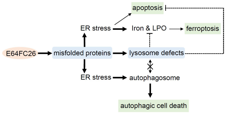
This study demonstrated that the PDI inhibitor, E64FC26, might induce ER stress and disrupt proteostasis through hindering the proper folding of proteins, and subsequently lead to lysosome defects. Defective lysosomes restricted the extent of apoptosis and ferroptosis induced by E64FC26-caused ER stress. Furthermore, defective lysosomes failed to form autolysosomes with autophagosomes, ultimately causing autophagic cell death of PDAC cells (Figure 8).
这项研究表明,PDI 抑制剂 E64FC26 可能通过阻碍蛋白质的正确折叠来诱导 ER 应激并破坏蛋白质稳态,进而导致溶酶体缺陷。有缺陷的溶酶体限制了 E64FC26 引起的 ER 应激诱导的细胞凋亡和铁死亡的程度。此外,有缺陷的溶酶体未能与自噬体形成自噬溶酶体,最终导致 PDAC 细胞自噬细胞死亡 ( Figure 8 )。
Figure 8. 图 8.
Possible mechanisms of E64FC26-induced cell death in pancreatic ductal adenocarcinoma (PDAC) cells. E64FC26 first induced endoplasmic reticular (ER) stress through impeding proper folding of proteins, and then subsequently caused lysosome defects. ER stress might have initiated ferroptosis and autophagy signals, and cells may have undergone partial apoptosis due to persistent ER stress from higher E64FC26 concentrations. However, defective lysosomes were unable to assist in the apoptosis and ferroptosis processes and hindered fusion with autophagosomes to form autolysosomes. Finally, E64FC26 only induced limited apoptosis and ferroptosis and mainly caused autophagic cell death. Solid lines are used to indicate activated pathways, and dashed lines are used to indicate inhibited pathways. LPO, lipid peroxidation.
胰腺导管腺癌 (PDAC) 细胞中 E64FC26 诱导细胞死亡的可能机制。E64FC26首先通过阻碍蛋白质的适当折叠诱导内质网 (ER) 应激,然后随后引起溶酶体缺陷。ER 应激可能引发了铁死亡和自噬信号,并且由于较高 ER 浓度的持续 ER 应激,细胞可能发生了部分凋亡E64FC26。然而,有缺陷的溶酶体无法协助细胞凋亡和铁死亡过程,并阻碍与自噬体融合形成自噬溶酶体。最后,E64FC26仅诱导有限的细胞凋亡和铁死亡,主要引起自噬细胞死亡。实线用于表示激活的通路,虚线用于表示受抑制的通路。LPO,脂质过氧化。
Acknowledgments 确认
We thank Anne Brunet (Department of Genetics, Stanford University, Stanford, CA, USA) for providing the FUW mCherry-GFP-LC3B plasmid.
我们感谢 Anne Brunet(美国加利福尼亚州斯坦福斯坦福大学遗传学系)提供 FUW mCherry-GFP-LC3B 质粒。
Author Contributions 作者贡献
Conceptualization: C.-S.H., W.-J.H. and Y.-C.L.; Data curation, C.-S.H., K.-L.L. and F.-H.S.; Formal analysis, C.-S.H., W.-J.H. and Y.-C.L.; Investigation, K.-L.L., W.-J.H., F.-H.S. and Y.-C.L.; Methodology, K.-L.L. and F.-H.S.; Validation, Y.-C.L.; Writing—original draft. C.-S.H. and Y.-C.L.; Writing—review and editing, Y.-C.L.; Funding acquisition, C.-S.H. and Y.-C.L. All authors have read and agreed to the published version of the manuscript.
概念化:C.-S.H.、W.-J.H.和 Y.-C.L.;数据管理,C.-S.H.,K.-L.L.和 F.-H.S.;形式分析,C.-S.H.,W.-J.H.和 Y.-C.L.;调查, K.-L.L., W.-J.H., F.-H.S.和 Y.-C.L.;方法论,K.-L.L.和 F.-H.S.;验证,Y.-C.L.;Writing - 原始草稿。C.-S.H. 和 Y.-C.L.;写作——审查和编辑,Y.-C.L.;资金收购,C.-S.H.和 Y.-C.L.所有作者均已阅读并同意手稿的已发表版本。
Institutional Review Board Statement
机构审查委员会声明
Not applicable. 不適用。
Informed Consent Statement
知情同意书
Not applicable. 不適用。
Data Availability Statement
数据可用性声明
The data are contained within the article.
数据包含在文章中。
Conflicts of Interest 利益冲突
The authors declare no conflict of interest.
作者声明没有利益冲突。
Funding Statement 资助声明
This research was funded by Ministry of Science and Technology of the Republic of China, grant numbers MOST 107-2320-B-038-023-MY3 and MOST 110-2320-B-038-061- and Wan Fang Hospital, grant numbers 111TMU-WFH-22 and 112TMU-WFH-13.
这项研究由中华民国科学技术部资助,资助号为 MOST 107-2320-B-038-023-MY3 和 MOST 110-2320-B-038-061- 和万方医院,资助号为 111TMU-WFH-22 和 112TMU-WFH-13。
Footnotes 脚注
Disclaimer/Publisher’s Note: The statements, opinions and data contained in all publications are solely those of the individual author(s) and contributor(s) and not of MDPI and/or the editor(s). MDPI and/or the editor(s) disclaim responsibility for any injury to people or property resulting from any ideas, methods, instructions or products referred to in the content.
免责声明/出版商注:所有出版物中包含的声明、意见和数据仅代表个人作者和贡献者的声明、意见和数据,与 MDPI 和/或编辑无关。MDPI 和/或编辑不对因内容中提及的任何想法、方法、说明或产品而造成的任何人员或财产伤害负责。
References 引用
-
1.Lukas J., Pospech J., Oppermann C., Hund C., Iwanov K., Pantoom S., Petters J., Frech M., Seemann S., Thiel F.G., et al. Role of endoplasmic reticulum stress and protein misfolding in disorders of the liver and pancreas. Adv. Med. Sci. 2019;64:315–323. doi: 10.1016/j.advms.2019.03.004IF: 2.5 Q3 . [DOI] [PubMed] [Google Scholar]
1.Lukas J., Pospech J., Oppermann C., Hund C., Iwanov K., Pantoom S., Petters J., Frech M., Seemann S., Thiel F.G., et al. 内质网应激和蛋白质错误折叠在肝脏和胰腺疾病中的作用。Adv. Med. Sci. 2019;64:315–323.doi: 10.1016/j.advms.2019.03.004IF: 2.5 Q3 .[ DOI ] [ PubMed ] [ Google Scholar ] [ ] -
2.Bulleid N.J., Ellgaard L. Multiple ways to make disulfides. Trends Biochem. Sci. 2011;36:485–492. doi: 10.1016/j.tibs.2011.05.004IF: 11.6 Q1 . [DOI] [PubMed] [Google Scholar]
2.Bulleid NJ., Ellgaard L. 制造二硫化物的多种方法。趋势生物化学。科学 2011;36:485–492.doi: 10.1016/j.tibs.2011.05.004IF: 11.6 Q1 .[ DOI ] [ PubMed ] [ Google Scholar ] [ ] -
3.Shergalis A.G., Hu S., Bankhead A., 3rd, Neamati N. Role of the ERO1-PDI interaction in oxidative protein folding and disease. Pharmacol. Ther. 2020;210:107525. doi: 10.1016/j.pharmthera.2020.107525IF: 12.0 Q1 . [DOI] [PMC free article] [PubMed] [Google Scholar]
3.Shergalis A.G., 胡 S., Bankhead A., 3rd, Neamati N. ERO1-PDI 相互作用在氧化蛋白折叠和疾病中的作用。药理学。Ther.2020;210:107525.doi: 10.1016/j.pharmthera.2020.107525IF: 12.0 Q1 .[ DOI ] [ PMC free article ] [ PubMed ] [ Google Scholar ] [ ] [ ] -
4.Xu X., Chiu J., Chen S., Fang C. Pathophysiological roles of cell surface and extracellular protein disulfide isomerase and their molecular mechanisms. Br. J. Pharmacol. 2021;178:2911–2930. doi: 10.1111/bph.15493IF: 6.8 Q1 . [DOI] [PubMed] [Google Scholar]
4.Xu X., Chiu J., Chen S., Fang C. 细胞表面和细胞外蛋白二硫键异构酶的病理生理作用及其分子机制。Br. J. 药理学杂志。2021 年;178:2911–2930.doi: 10.1111/bph.15493IF: 6.8 Q1 .[ DOI ] [ PubMed ] [ Google Scholar ] [ ] -
5.Zhang Z., Zhang L., Zhou L., Lei Y., Zhang Y., Huang C. Redox signaling and unfolded protein response coordinate cell fate decisions under ER stress. Redox Biol. 2019;25:101047. doi: 10.1016/j.redox.2018.11.005IF: 10.7 Q1 . [DOI] [PMC free article] [PubMed] [Google Scholar]
5.Zhang Z., Zhang L., 周 L., Lei Y., Zhang Y., Huang C. 氧化还原信号传导和未折叠的蛋白质反应协调 ER 应激下的细胞命运决定。氧化还原生物学 2019;25:101047。doi: 10.1016/j.redox.2018.11.005IF: 10.7 Q1 .[ DOI ] [ PMC free article ] [ PubMed ] [ Google Scholar ] [ ] [ ] -
6.Jha V., Kumari T., Manickam V., Assar Z., Olson K.L., Min J.K., Cho J. ERO1-PDI Redox Signaling in Health and Disease. Antioxid. Redox Signal. 2021;35:1093–1115. doi: 10.1089/ars.2021.0018IF: 5.9 Q1 . [DOI] [PMC free article] [PubMed] [Google Scholar]
6.Jha V.、Kumari T.、Manickam V.、Assar Z.、Olson KL、Min JK、Cho J. 健康和疾病中的 ERO1-PDI 氧化还原信号传导。抗氧化。氧化还原信号。2021;35:1093–1115.doi: 10.1089/ars.2021.0018IF: 5.9 Q1 .[ DOI ] [ PMC free article ] [ PubMed ] [ Google Scholar ] [ ] [ ] -
7.Stepanchuk A.A., Stys P.K. Amyloid dye pairs as spectral sensors for enhanced detection and differentiation of misfolded proteins. J. Photochem. Photobiol. B. 2023;248:112786. doi: 10.1016/j.jphotobiol.2023.112786IF: 3.9 Q1 . [DOI] [PubMed] [Google Scholar]
7.Stepanchuk A.A., Stys P.K. 淀粉样蛋白染料对作为增强错误折叠蛋白质检测和区分的光谱传感器。光生物学。B. 2023;248:112786。doi: 10.1016/j.jphotobiol.2023.112786IF: 3.9 Q1 .[ DOI ] [ PubMed ] [ Google Scholar ] [ ] -
8.Yamada Y., Tomaru U., Ishizu A., Ito T., Kiuchi T., Ono A., Miyajima S., Nagai K., Higashi T., Matsuno Y., et al. Decreased proteasomal function accelerates cigarette smoke-induced pulmonary emphysema in mice. Lab. Investig. 2015;95:625–634. doi: 10.1038/labinvest.2015.43IF: 5.1 Q1 . [DOI] [PubMed] [Google Scholar]
8.Yamada Y., Tomaru U., Ishizu A., Ito T., Kiuchi T., Ono A., Miyajima S., Nagai K., Higashi T., Matsuno Y., et al. 蛋白酶体功能下降加速香烟烟雾诱导的小鼠肺气肿。实验室调查 2015;95:625–634.doi: 10.1038/labinvest.2015.43IF: 5.1 Q1 .[ DOI ] [ PubMed ] [ Google Scholar ] [ ] -
9.Brusa I., Sondo E., Falchi F., Pedemonte N., Roberti M., Cavalli A. Proteostasis Regulators in Cystic Fibrosis: Current Development and Future Perspectives. J. Med. Chem. 2022;65:5212–5243. doi: 10.1021/acs.jmedchem.1c01897IF: 6.8 Q1 . [DOI] [PMC free article] [PubMed] [Google Scholar]
9.Brusa I., Sondo E., Falchi F., Pedemonte N., Roberti M., Cavalli A. 囊性纤维化中的蛋白质稳态调节剂:当前发展和未来前景。J. Med. Chem. 2022;65:5212–5243.doi: 10.1021/acs.jmedchem.1c01897IF: 6.8 Q1 .[ DOI ] [ PMC free article ] [ PubMed ] [ Google Scholar ] [ ] [ ] -
10.Ye Z.W., Zhang J., Aslam M., Blumental-Perry A., Tew K.D., Townsend D.M. Protein disulfide isomerase family mediated redox regulation in cancer. Adv. Cancer Res. 2023;160:83–106. doi: 10.1016/bs.acr.2023.06.001. [DOI] [PMC free article] [PubMed] [Google Scholar]
10.Ye Z.W., Zhang J., Aslam M., Blumental-Perry A., Tew KD, Townsend DM 蛋白质二硫键异构酶家族介导的癌症氧化还原调节。癌症研究 2023 年;160:83–106.doi: 10.1016/bs.acr.2023.06.001.[ DOI ] [ PMC free article ] [ PubMed ] [ Google Scholar ] [ ] [ ] -
11.Hoffstrom B.G., Kaplan A., Letso R., Schmid R.S., Turmel G.J., Lo D.C., Stockwell B.R. Inhibitors of protein disulfide isomerase suppress apoptosis induced by misfolded proteins. Nat. Chem. Biol. 2010;6:900–906. doi: 10.1038/nchembio.467IF: 12.9 Q1 . [DOI] [PMC free article] [PubMed] [Google Scholar]
11.Hoffstrom B.G., Kaplan A., Letso R., Schmid R.S., Turmel G.J., Lo D.C., Stockwell B.R. 蛋白质二硫键异构酶抑制剂抑制错误折叠蛋白质诱导的细胞凋亡。Nat. Chem. Biol. 2010;6:900–906.doi: 10.1038/nchembio.467IF: 12.9 Q1 .[ DOI ] [ PMC free article ] [ PubMed ] [ Google Scholar ] [ ] [ ] -
12.Wise R., Duhachek-Muggy S., Qi Y., Zolkiewski M., Zolkiewska A. Protein disulfide isomerases in the endoplasmic reticulum promote anchorage-independent growth of breast cancer cells. Breast Cancer Res. Treat. 2016;157:241–252. doi: 10.1007/s10549-016-3820-1IF: 3.0 Q2 . [DOI] [PMC free article] [PubMed] [Google Scholar]
12.Wise R., Duhachek-Muggy S., Qi Y., Zolkiewski M., Zolkiewska A. 内质网中的蛋白质二硫键异构酶促进乳腺癌细胞不依赖锚定的生长。乳腺癌研究治疗。2016;157:241–252.doi: 10.1007/s10549-016-3820-1IF: 3.0 Q2 .[ DOI ] [ PMC free article ] [ PubMed ] [ Google Scholar ] [ ] [ ] -
13.Ramos F.S., Serino L.T., Carvalho C.M., Lima R.S., Urban C.A., Cavalli I.J., Ribeiro E.M. PDIA3 and PDIA6 gene expression as an aggressiveness marker in primary ductal breast cancer. Genet. Mol. Res. 2015;14:6960–6967. doi: 10.4238/2015.June.26.4IF: 0.6 Q4 . [DOI] [PubMed] [Google Scholar]
13.Ramos FS, Serino L.T., Carvalho CM, Lima RS, Urban C.A., Cavalli IJ, Ribeiro EM PDIA3 和 PDIA6 基因表达作为原发性导管乳腺癌的侵袭性标志物。基因。Mol. Res. 2015;14:6960–6967.doi: 10.4238/2015.June.26.4IF: 0.6 Q4 .[ DOI ] [ PubMed ] [ Google Scholar ] [ ] -
14.Alavi M., Mah V., Maresh E.L., Bagryanova L., Horvath S., Chia D., Goodglick L., Liu A.Y. High expression of AGR2 in lung cancer is predictive of poor survival. BMC Cancer. 2015;15:655. doi: 10.1186/s12885-015-1658-2IF: 3.4 Q2 . [DOI] [PMC free article] [PubMed] [Google Scholar]
14.Alavi M., Mah V., Maresh E.L., Bagryanova L., Horvath S., Chia D., Goodglick L., Liu A.Y. AGR2 在肺癌中的高表达预示着生存率低。BMC 癌症。2015;15:655.doi: 10.1186/s12885-015-1658-2IF: 3.4 Q2 .[ DOI ] [ PMC free article ] [ PubMed ] [ Google Scholar ] [ ] [ ] -
15.Mouawad R., Neamati N. Inhibition of Protein Disulfide Isomerase (PDIA1) Leads to Proteasome-Mediated Degradation of Ubiquitin-like PHD and RING Finger Domain-Containing Protein 1 (UHRF1) and Increased Sensitivity of Glioblastoma Cells to Topoisomerase II Inhibitors. ACS Pharmacol. Transl. Sci. 2023;6:100–114. doi: 10.1021/acsptsci.2c00186IF: 4.9 Q1 . [DOI] [PMC free article] [PubMed] [Google Scholar]
15.Mouawad R., Neamati N. 抑制蛋白二硫键异构酶 (PDIA1) 导致蛋白酶体介导的泛素样 PHD 和含无名指结构域的蛋白 1 (UHRF1) 的降解,并增加胶质母细胞瘤细胞对拓扑异构酶 II 抑制剂的敏感性。ACS 药理学。Transl. Sci. 2023;6:100–114.doi: 10.1021/acsptsci.2c00186IF: 4.9 Q1 .[ DOI ] [ PMC free article ] [ PubMed ] [ Google Scholar ] [ ] [ ] -
16.Araujo T.L.S., Zeidler J.D., Oliveira P.V.S., Dias M.H., Armelin H.A., Laurindo F.R.M. Protein disulfide isomerase externalization in endothelial cells follows classical and unconventional routes. Free Radic. Biol. Med. 2017;103:199–208. doi: 10.1016/j.freeradbiomed.2016.12.021IF: 7.1 Q1 . [DOI] [PubMed] [Google Scholar]
16.Araujo T.L.S., Zeidler J.D., Oliveira P.V.S., Dias M.H., Armelin H.A., Laurindo F.R.M. 内皮细胞中的蛋白质二硫键异构酶外化遵循经典和非常规途径。自由 Radic。生物医学 2017;103:199–208.doi: 10.1016/j.freeradbiomed.2016.12.021IF: 7.1 Q1 .[ DOI ] [ PubMed ] [ Google Scholar ] [ ] -
17.Wolzak K., Vermunt L., Campo M.D., Jorge-Oliva M., van Ziel A.M., Li K.W., Smit A.B., Chen-Ploktkin A., Irwin D.J., Lemstra A.W., et al. Protein disulfide isomerases as CSF biomarkers for the neuronal response to tau pathology. Alzheimers Dement. 2023;19:3563–3574. doi: 10.1002/alz.12978IF: 13.0 Q1 . [DOI] [PubMed] [Google Scholar]
17.Wolzak K., Vermunt L., Campo M.D., Jorge-Oliva M., van Ziel A.M., Li K.W., Smit A.B., Chen-Ploktkin A., Irwin D.J., Lemstra A.W., et al. 蛋白二硫键异构酶作为神经元对 tau 病理反应的 CSF 生物标志物。阿尔茨海默病 Dement。2023;19:3563–3574.doi: 10.1002/alz.12978IF: 13.0 Q1 .[ DOI ] [ PubMed ] [ Google Scholar ] [ ] -
18.Jang I., Pottekat A., Poothong J., Yong J., Lagunas-Acosta J., Charbono A., Chen Z., Scheuner D.L., Liu M., Itkin-Ansari P., et al. PDIA1/P4HB is required for efficient proinsulin maturation and ß cell health in response to diet induced obesity. eLife. 2019;8:e44528. doi: 10.7554/eLife.44528IF: 6.4 Q1 . [DOI] [PMC free article] [PubMed] [Google Scholar]
18.Jang I., Pottekat A., Poothong J., Yong J., Lagunas-Acosta J., Charbono A., Chen Z., Scheuner D.L., Liu M., Itkin-Ansari P., et al. PDIA1/P4HB 是响应饮食诱导的肥胖的有效胰岛素原成熟和 ß 细胞健康所必需的。电子生活。2019 年;8:e44528.doi: 10.7554/eLife.44528IF: 6.4 Q1 .[ DOI ] [ PMC free article ] [ PubMed ] [ Google Scholar ] [ ] [ ] -
19.Xu S., Butkevich A.N., Yamada R., Zhou Y., Debnath B., Duncan R., Zandi E., Petasis N.A., Neamati N. Discovery of an orally active small-molecule irreversible inhibitor of protein disulfide isomerase for ovarian cancer treatment. Proc. Natl. Acad. Sci. USA. 2012;109:16348–16353. doi: 10.1073/pnas.1205226109IF: 9.4 Q1 . [DOI] [PMC free article] [PubMed] [Google Scholar]
19.徐S., 布特克维奇A.N., 山田R., 周Y., Debnath B., Duncan R., Zandi E., Petasis N.A., Neamati N. 发现一种口服活性小分子不可逆蛋白二硫键异构酶抑制剂治疗卵巢癌。美国国家科学院院刊,2012 年;109:16348–16353.doi: 10.1073/pnas.1205226109IF: 9.4 Q1 .[ DOI ] [ PMC free article ] [ PubMed ] [ Google Scholar ] [ ] [ ] -
20.Powell L.E., Foster P.A. Protein disulphide isomerase inhibition as a potential cancer therapeutic strategy. Cancer Med. 2021;10:2812–2825. doi: 10.1002/cam4.3836IF: 2.9 Q2 . [DOI] [PMC free article] [PubMed] [Google Scholar]
20.Powell L.E., Foster P.A. 蛋白质二硫化物异构酶抑制作为一种潜在的癌症治疗策略。癌症医学 2021;10:2812–2825.doi: 10.1002/cam4.3836IF: 2.9 Q2 .[ DOI ] [ PMC free article ] [ PubMed ] [ Google Scholar ] [ ] [ ] -
21.Vatolin S., Phillips J.G., Jha B.K., Govindgari S., Hu J., Grabowski D., Parker Y., Lindner D.J., Zhong F., Distelhorst C.W., et al. Novel Protein Disulfide Isomerase Inhibitor with Anticancer Activity in Multiple Myeloma. Cancer Res. 2016;76:3340–3350. doi: 10.1158/0008-5472.CAN-15-3099IF: 12.5 Q1 . [DOI] [PubMed] [Google Scholar]
21.瓦托林 S., 菲利普斯 J.G., Jha B.K., 戈文德加里 S., 胡 J., 格拉博夫斯基 D., 帕克 Y., 林德纳 D.J., 钟 F., 迪斯特尔霍斯特 C.W., 等. 多发性骨髓瘤中具有抗癌活性的新型蛋白二硫键异构酶抑制剂.癌症研究 2016 年;76:3340–3350.doi: 10.1158/0008-5472.CAN-15-3099IF: 12.5 Q1 .[ DOI ] [ PubMed ] [ Google Scholar ] [ ] -
22.Robinson R.M., Reyes L., Duncan R.M., Bian H., Reitz A.B., Manevich Y., McClure J.J., Champion M.M., Chou C.J., Sharik M.E., et al. Inhibitors of the protein disulfide isomerase family for the treatment of multiple myeloma. Leukemia. 2019;33:1011–1022. doi: 10.1038/s41375-018-0263-1IF: 12.8 Q1 . [DOI] [PMC free article] [PubMed] [Google Scholar]
22.Robinson R.M., Reyes L., Duncan R.M., Bian H., Reitz A.B., Manevich Y., McClure J.J., Champion M.M., Chou C.J., Sharik M.E., et al. 蛋白二硫键异构酶家族抑制剂,用于治疗多发性骨髓瘤。白血病。2019;33:1011–1022.doi: 10.1038/s41375-018-0263-1IF: 12.8 Q1 .[ DOI ] [ PMC free article ] [ PubMed ] [ Google Scholar ] [ ] [ ] -
23.Zhou X., Li G., Kaplan A., Gaschler M.M., Zhang X., Hou Z., Jiang M., Zott R., Cremers S., Stockwell B.R., et al. Small molecule modulator of protein disulfide isomerase attenuates mutant huntingtin toxicity and inhibits endoplasmic reticulum stress in a mouse model of Huntington’s disease. Hum. Mol. Genet. 2018;27:1545–1555. doi: 10.1093/hmg/ddy061IF: 3.1 Q2 . [DOI] [PMC free article] [PubMed] [Google Scholar]
23.周晓明, 李国, 卡普兰A., 加施勒M.M., 张晓明, 侯志强, 江明明, 佐特R., Cremers S., 斯托克韦尔B.R., 等. 蛋白二硫键异构酶的小分子调节剂在亨廷顿病小鼠模型中减弱突变型亨廷顿蛋白毒性并抑制内质网应激。Hum. Mol. Genet. 2018;27:1545–1555.doi: 10.1093/hmg/ddy061IF: 3.1 Q2 .[ DOI ] [ PMC free article ] [ PubMed ] [ Google Scholar ] [ ] [ ] - 24.Kyani A., Tamura S., Yang S., Shergalis A., Samanta S., Kuang Y., Ljungman M., Neamati N. Discovery and Mechanistic Elucidation of a Class of Protein Disulfide Isomerase Inhibitors for the Treatment of Glioblastoma. ChemMedChem. 2018;13:164–177. doi: 10.1002/cmdc.201700629IF: 3.6 Q2 . [DOI] [PMC free article] [PubMed] [Google Scholar]
- 25.Tang Z., Kang B., Li C., Chen T., Zhang Z. GEPIA2: An enhanced web server for large-scale expression profiling and interactive analysis. Nucleic Acids Res. 2019;47:W556–W560. doi: 10.1093/nar/gkz430IF: 16.6 Q1 . [DOI] [PMC free article] [PubMed] [Google Scholar]
- 26.Yu J., Li T., Liu Y., Wang X., Zhang J., Wang X., Shi G., Lou J., Wang L., Wang C.C., et al. Phosphorylation switches protein disulfide isomerase activity to maintain proteostasis and attenuate ER stress. EMBO J. 2020;39:e103841. doi: 10.15252/embj.2019103841IF: 9.4 Q1 . [DOI] [PMC free article] [PubMed] [Google Scholar]
- 27.Hurst K.E., Lawrence K.A., Reyes Angeles L., Ye Z., Zhang J., Townsend D.M., Dolloff N., Thaxton J.E. Endoplasmic Reticulum Protein Disulfide Isomerase Shapes T Cell Efficacy for Adoptive Cellular Therapy of Tumors. Cells. 2019;8:1514. doi: 10.3390/cells8121514IF: 5.1 Q2 . [DOI] [PMC free article] [PubMed] [Google Scholar]
- 28.Yang W.S., Stockwell B.R. Ferroptosis: Death by Lipid Peroxidation. Trends Cell Biol. 2016;26:165–176. doi: 10.1016/j.tcb.2015.10.014IF: 13.0 Q1 . [DOI] [PMC free article] [PubMed] [Google Scholar]
- 29.Torii S., Shintoku R., Kubota C., Yaegashi M., Torii R., Sasaki M., Suzuki T., Mori M., Yoshimoto Y., Takeuchi T., et al. An essential role for functional lysosomes in ferroptosis of cancer cells. Biochem. J. 2016;473:769–777. doi: 10.1042/BJ20150658IF: 4.4 Q2 . [DOI] [PubMed] [Google Scholar]
- 30.Piffoux M., Eriau E., Cassier P.A. Autophagy as a therapeutic target in pancreatic cancer. Br. J. Cancer. 2021;124:333–344. doi: 10.1038/s41416-020-01039-5IF: 6.4 Q1 . [DOI] [PMC free article] [PubMed] [Google Scholar]
- 31.Yang S., Wang X., Contino G., Liesa M., Sahin E., Ying H., Bause A., Li Y., Stommel J.M., Dell’antonio G., et al. Pancreatic cancers require autophagy for tumor growth. Genes. Dev. 2011;25:717–729. doi: 10.1101/gad.2016111IF: 7.5 Q1 . [DOI] [PMC free article] [PubMed] [Google Scholar]
- 32.Rahman N.S.A., Zahari S., Syafruddin S.E., Firdaus-Raih M., Low T.Y., Mohtar M.A. Functions and mechanisms of protein disulfide isomerase family in cancer emergence. Cell Biosci. 2022;12:129. doi: 10.1186/s13578-022-00868-6IF: 6.1 Q1 . [DOI] [PMC free article] [PubMed] [Google Scholar]
- 33.Khodier C., VerPlank L., Nag P.P., Pu J., Wurst J., Pilyugina T., Dockendorff C., Galinski C.N., Scalise A.A., Passam F., et al. Probe Reports from the NIH Molecular Libraries Program. National Center for Biotechnology Information (US); Bethesda, MD, USA: 2010. Identification of ML359 as a Small Molecule Inhibitor of Protein Disulfide Isomerase. [PubMed] [Google Scholar]
- 34.Kaplan A., Gaschler M.M., Dunn D.E., Colligan R., Brown L.M., Palmer A.G., 3rd, Lo D.C., Stockwell B.R. Small molecule-induced oxidation of protein disulfide isomerase is neuroprotective. Proc. Natl. Acad. Sci. USA. 2015;112:E2245–E2252. doi: 10.1073/pnas.1500439112IF: 9.4 Q1 . [DOI] [PMC free article] [PubMed] [Google Scholar]
- 35.Lin L., Gopal S., Sharda A., Passam F., Bowley S.R., Stopa J., Xue G., Yuan C., Furie B.C., Flaumenhaft R., et al. Quercetin-3-rutinoside Inhibits Protein Disulfide Isomerase by Binding to Its b’x Domain. J. Biol. Chem. 2015;290:23543–23552. doi: 10.1074/jbc.M115.666180IF: 4.0 Q2 . [DOI] [PMC free article] [PubMed] [Google Scholar]
- 36.Cole K.S., Grandjean J.M.D., Chen K., Witt C.H., O’Day J., Shoulders M.D., Wiseman R.L., Weerapana E. Characterization of an A-Site Selective Protein Disulfide Isomerase A1 Inhibitor. Biochemistry. 2018;57:2035–2043. doi: 10.1021/acs.biochem.8b00178IF: 2.9 Q3 . [DOI] [PMC free article] [PubMed] [Google Scholar]
- 37.Alfieri M.L., Cariola A., Panzella L., Napolitano A., d’Ischia M., Valgimigli L., Crescenzi O. Disentangling the Puzzling Regiochemistry of Thiol Addition to o-Quinones. J. Org. Chem. 2022;87:4580–4589. doi: 10.1021/acs.joc.1c02911IF: 3.3 Q1 . [DOI] [PMC free article] [PubMed] [Google Scholar]
- 38.Robinson R.M., Reyes L., Duncan R.M., Bian H., Strobel E.D., Hyman S.L., Reitz A.B., Dolloff N.G. Tuning isoform selectivity and bortezomib sensitivity with a new class of alkenyl indene PDI inhibitor. Eur. J. Med. Chem. 2020;186:111906. doi: 10.1016/j.ejmech.2019.111906IF: 6.0 Q1 . [DOI] [PMC free article] [PubMed] [Google Scholar]
- 39.Deer E.L., González-Hernández J., Coursen J.D., Shea J.E., Ngatia J., Scaife C.L., Firpo M.A., Mulvihill S.J. Phenotype and genotype of pancreatic cancer cell lines. Pancreas. 2010;39:425–435. doi: 10.1097/MPA.0b013e3181c15963IF: 1.7 Q3 . [DOI] [PMC free article] [PubMed] [Google Scholar]
- 40.Sawai H., Funahashi H., Yamamoto M., Okada Y., Hayakawa T., Tanaka M., Takeyama H., Manabe T. Interleukin-1alpha enhances integrin alpha(6)beta(1) expression and metastatic capability of human pancreatic cancer. Oncology. 2003;65:167–173. doi: 10.1159/000072343IF: 2.5 Q3 . [DOI] [PubMed] [Google Scholar]
- 41.Zeng S., Pöttler M., Lan B., Grützmann R., Pilarsky C., Yang H. Chemoresistance in Pancreatic Cancer. Int. J. Mol. Sci. 2019;20:4504. doi: 10.3390/ijms20184504IF: 4.9 Q1 . [DOI] [PMC free article] [PubMed] [Google Scholar]
- 42.Zhao B., Qin C., Li Z., Wang Y., Li T., Cao H., Yang X., Li T., Wang W. Multidrug resistance genes screening of pancreatic ductal adenocarcinoma based on sensitivity profile to chemotherapeutic drugs. Cancer Cell Int. 2022;22:374. doi: 10.1186/s12935-022-02785-7IF: 5.3 Q1 . [DOI] [PMC free article] [PubMed] [Google Scholar]
- 43.Zhu S., Zhang Q., Sun X., Zeh H.J., 3rd, Lotze M.T., Kang R., Tang D. HSPA5 Regulates Ferroptotic Cell Death in Cancer Cells. Cancer Res. 2017;77:2064–2077. doi: 10.1158/0008-5472.CAN-16-1979IF: 12.5 Q1 . [DOI] [PMC free article] [PubMed] [Google Scholar]
- 44.Fu X., Cui J., Meng X., Jiang P., Zheng Q., Zhao W., Chen X. Endoplasmic reticulum stress, cell death and tumor: Association between endoplasmic reticulum stress and the apoptosis pathway in tumors (Review) Oncol. Rep. 2021;45:801–808. doi: 10.3892/or.2021.7933IF: 3.8 Q2 . [DOI] [PMC free article] [PubMed] [Google Scholar]
- 45.Kuang H., Sun X., Liu Y., Tang M., Wei Y., Shi Y., Li R., Xiao G., Kang J., Wang F., et al. Palmitic acid-induced ferroptosis via CD36 activates ER stress to break calcium-iron balance in colon cancer cells. FEBS J. 2023;290:3664–3687. doi: 10.1111/febs.16772IF: 5.5 Q1 . [DOI] [PubMed] [Google Scholar]
- 46.Park E.J., Park Y.J., Lee S.J., Lee K., Yoon C. Whole cigarette smoke condensates induce ferroptosis in human bronchial epithelial cells. Toxicol. Lett. 2019;303:55–66. doi: 10.1016/j.toxlet.2018.12.007IF: 2.9 Q2 . [DOI] [PubMed] [Google Scholar]
- 47.Gao H., Bai Y., Jia Y., Zhao Y., Kang R., Tang D., Dai E. Ferroptosis is a lysosomal cell death process. Biochem. Biophys. Res. Commun. 2018;503:1550–1556. doi: 10.1016/j.bbrc.2018.07.078IF: 2.5 Q3 . [DOI] [PubMed] [Google Scholar]
- 48.Yagoda N., von Rechenberg M., Zaganjor E., Bauer A.J., Yang W.S., Fridman D.J., Wolpaw A.J., Smukste I., Peltier J.M., Boniface J.J., et al. RAS-RAF-MEK-dependent oxidative cell death involving voltage-dependent anion channels. Nature. 2007;447:864–868. doi: 10.1038/nature05859IF: 50.5 Q1 . [DOI] [PMC free article] [PubMed] [Google Scholar]
- 49.Sun K., Li C., Liao S., Yao X., Ouyang Y., Liu Y., Wang Z., Li Z., Yao F. Ferritinophagy, a form of autophagic ferroptosis: New insights into cancer treatment. Front. Pharmacol. 2022;13:1043344. doi: 10.3389/fphar.2022.1043344IF: 4.4 Q1 . [DOI] [PMC free article] [PubMed] [Google Scholar]
- 50.Koksal A.R., Verne G.N., Zhou Q. Endoplasmic reticulum stress in biological processing and disease. J. Investig. Med. 2021;69:309–315. doi: 10.1136/jim-2020-001570IF: 2.5 Q1 . [DOI] [PubMed] [Google Scholar]
- 51.Yuan W., Fang W., Zhang R., Lyu H., Xiao S., Guo D., Ali D.W., Michalak M., Chen X.Z., Zhou C., et al. Therapeutic strategies targeting AMPK-dependent autophagy in cancer cells. Biochim. Biophys. Acta Mol. Cell Res. 2023;1870:119537. doi: 10.1016/j.bbamcr.2023.119537IF: 4.6 Q1 . [DOI] [PubMed] [Google Scholar]
- 52.Hubert V., Weiss S., Rees A.J., Kain R. Modulating Chaperone-Mediated Autophagy and Its Clinical Applications in Cancer. Cells. 2022;11:2562. doi: 10.3390/cells11162562IF: 5.1 Q2 . [DOI] [PMC free article] [PubMed] [Google Scholar]
- 53.Kang R., Tang D. Autophagy and Ferroptosis—What’s the Connection? Curr. Pathobiol. Rep. 2017;5:153–159. doi: 10.1007/s40139-017-0139-5. [DOI] [PMC free article] [PubMed] [Google Scholar]
- 54.Ogata M., Hino S., Saito A., Morikawa K., Kondo S., Kanemoto S., Murakami T., Taniguchi M., Tanii I., Yoshinaga K., et al. Autophagy is activated for cell survival after endoplasmic reticulum stress. Mol. Cell Biol. 2006;26:9220–9231. doi: 10.1128/MCB.01453-06IF: 3.2 Q2 . [DOI] [PMC free article] [PubMed] [Google Scholar]
- 55.Patel S., Homaei A., El-Seedi H.R., Akhtar N. Cathepsins: Proteases that are vital for survival but can also be fatal. Biomed. Pharmacother. 2018;105:526–532. doi: 10.1016/j.biopha.2018.05.148IF: 6.9 Q1 . [DOI] [PMC free article] [PubMed] [Google Scholar]
- 56.Turk V., Stoka V., Vasiljeva O., Renko M., Sun T., Turk B., Turk D. Cysteine cathepsins: From structure, function and regulation to new frontiers. Biochim. Biophys. Acta. 2012;1824:68–88. doi: 10.1016/j.bbapap.2011.10.002IF: 2.5 Q3 . [DOI] [PMC free article] [PubMed] [Google Scholar]
- 57.Suk F.M., Chang C.C., Sun P.C., Ke W.T., Chung C.C., Lee K.L., Chan T.S., Liang Y.C. MCPIP1 Enhances TNF-α-Mediated Apoptosis through Downregulation of the NF-κB/cFLIP Axis. Biology. 2021;10:655. doi: 10.3390/biology10070655IF: 3.6 Q1 . [DOI] [PMC free article] [PubMed] [Google Scholar]
- 58.Chang C.C., Lee K.L., Chan T.S., Chung C.C., Liang Y.C. Histone Deacetylase Inhibitors Downregulate Calcium Pyrophosphate Crystal Formation in Human Articular Chondrocytes. Int. J. Mol. Sci. 2022;23:2604. doi: 10.3390/ijms23052604IF: 4.9 Q1 . [DOI] [PMC free article] [PubMed] [Google Scholar]
- 59.Liang Y.C., Chang C.C., Sheu M.T., Lin S.Y., Chung C.C., Teng C.T., Suk F.M. The Antihistamine Deptropine Induces Hepatoma Cell Death through Blocking Autophagosome-Lysosome Fusion. Cancers. 2020;12:1610. doi: 10.3390/cancers12061610IF: 4.5 Q1 . [DOI] [PMC free article] [PubMed] [Google Scholar]
- 60.Thomé M.P., Filippi-Chiela E.C., Villodre E.S., Migliavaca C.B., Onzi G.R., Felipe K.B., Lenz G. Ratiometric analysis of Acridine Orange staining in the study of acidic organelles and autophagy. J. Cell Sci. 2016;129:4622–4632. doi: 10.1242/jcs.195057IF: 3.3 Q3 . [DOI] [PubMed] [Google Scholar]
Associated Data
This section collects any data citations, data availability statements, or supplementary materials included in this article.
Data Availability Statement
The data are contained within the article.


